However, many therapeutic and diagnostic agents in general may not be delivered using these routes because they might be susceptible to
enzymatic degradation or cannot be absorbed into the
systemic circulation efficiently due to
molecular size and charge issues, and thus, will not be fully therapeutically effective.
There are several known problems associated with the injection process.
One of such problems is undesirable
extravasation of the diagnostic or therapeutic agents into tissue, which is particularly prevalent with intravenously injected agents.
Extravasation generally refers to leakage of fluids out of a container, and more specifically refers to leakage of intravenous drugs from a
vein into surrounding tissues, resulting in an injury to the tissues.
Once the intravenous
extravasation has occurred, damage can continue for months and involve nerves, tendons and joints.
If treatment is delayed, surgical debridement,
skin grafting, and even
amputation have been known to be the unfortunate consequences.
Occurrence of extravasation is possible with all intravenuous drugs, but it is a particularly significant problem with cytoxic drugs used for treatment of
cancer (i.e. during
chemotherapy).
Unfortunately, it cannot tell the difference between a
cancer cell and some healthy cells.
With permanent implants (e.g.
prostate), the radioactivity of the seeds typically decays with time.
Veins of people receiving
chemotherapy are often fragile, mobile, and difficult to cannulate.
Patients who have had previous
radiation therapy at the site of injection may develop severe local reactions from cytotoxic drugs.
Cytotoxic drugs also have the potential to cause cutaneous abnormalities in areas that have been damaged previously by
radiation, even in areas that are distant from the
injection site.
Furthermore, areas of previous
surgery where the underlying tissue is likely to be fibrosed and toughened dramatically present an
increased risk of extravasation.
Some
chemotherapy drugs often never reach the tumors they are intended to treat because the blood vessels feeding the tumors are abnormal.
A tumor's capillaries (small blood vessels that directly deliver
oxygen and nutrients to
cancer cells) can be irregularly shaped, being excessively thin in some areas and forming thick, snarly clumps in others.
These malformations create a turbulent, uneven
blood flow, so that too much blood goes to one region of the tumor, and too little to another.
In addition, the
capillary endothelial cells lining the inner surface of tumor capillaries, normally a smooth, tightly-packed sheet, have gaps between them, causing vessel leakiness.
However, while some experimental uses of the localized delivery of cytotoxic drugs have been attempted, there has been little implementation of such
drug delivery in practice, possibly due to numerous problems associated with such delivery.
First, it is often necessary to deliver cytotoxic drugs to remote and not easily accessible blood vessels and other lumens within
body organs, such as lungs.
Moreover, the existing methods lack the ability to contain the cytotoxic agent and / or
radiation therapy and mitigate collateral damage to non-affected
anatomy and structures.
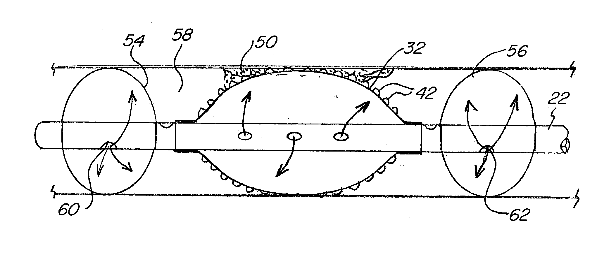
While the above described catheter devices are useful for delivering the drugs to a specific
target site, these systems are not particularly efficient at infusing the relevant biological material with the
drug.
Instead, the catheter may need to remain in place for an unnecessarily long period of time while the infusion of the drug into the biological material is allowed to take place.
This is undesirable, especially in applications such as pulmonology, where the patient's respiratory passage has been somewhat restricted by the device.
Further, this can result in some of the agent never being infused into the targeted material and instead remaining in the cavity and, after the
balloon catheter is removed, subsequently migrating to other undesired portions of the body.


