
Peptide homing to brain tumors

a brain tumor and peptide technology, applied in the field of peptides, can solve the problems of poor prognosis, poor treatment effect, and difficulty in detecting low-grade astrocytomas

Inactive Publication Date: 2011-06-02
BURNHAM INST FOR MEDICAL RES +1
View PDF3 Cites 4 Cited by
  • Summary
  • Abstract
  • Description
  • Claims
  • Application Information

AI Technical Summary

Benefits of technology

The present invention is about a new peptide called CGLSGLGVA, which specifically targets astrocytoma brain cancer cells. The peptide was identified using a phage display screen and has been shown to be effective in diagnosing and treating brain cancers. The invention provides a peptide composition that can be used for targeted therapy and diagnosis of brain cancers. The peptide can be administered to patients as a pharmaceutical composition or can be labeled and used for diagnosis. Overall, the invention provides a novel tool for brain cancer diagnosis and treatment.

Problems solved by technology

The prognosis for patients suffering from brain tumors is poor and has not improved during the last decades.
Especially low-grade astrocytomas are challenging because they have shown to be unreachable by conventional treatment strategies such as radiation or surgery.

Method used


Image

  • Peptide homing to brain tumors
  • Peptide homing to brain tumors
  • Peptide homing to brain tumors

Examples


example 1

Homing of the Phage Displaying Peptide Sequence CGLSGLGVA to the Intracranial Islet Tumors

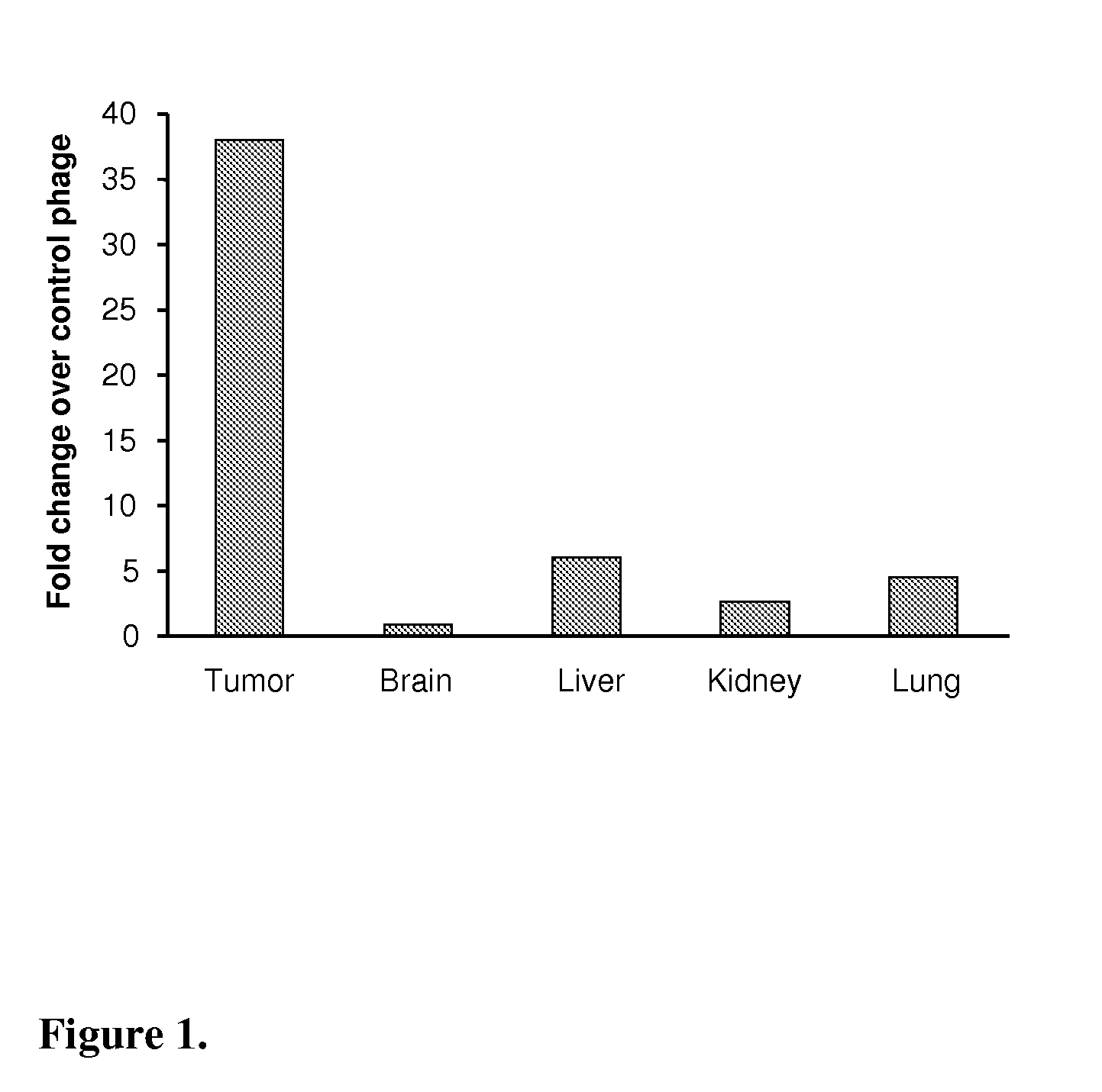
[0032]CGLSGLGVA or a control phage (109 pfu=plaque forming unit) were injected into the tail vein of intracranial HIF-1alfa KO (HIFko) tumor-bearing mice. After 15 min of circulation the mice were perfused through the heart with saline to remove unbound phage followed by excision of tumor containing brain, normal brain, liver, kidney and lungs. The number of phage homed to those organs were determined by titration. FIG. 1 shows as a bar graph that the peptide very specifically homes to the intracranial, early stage astrocytoma model (HIFko) that grows as islets and harbors co-opted tumor vessels in the brain.

example 2

Homing of the Fluorescein-Conjugated CGLSGLGVA Peptide to the Intracranial Islet Tumors

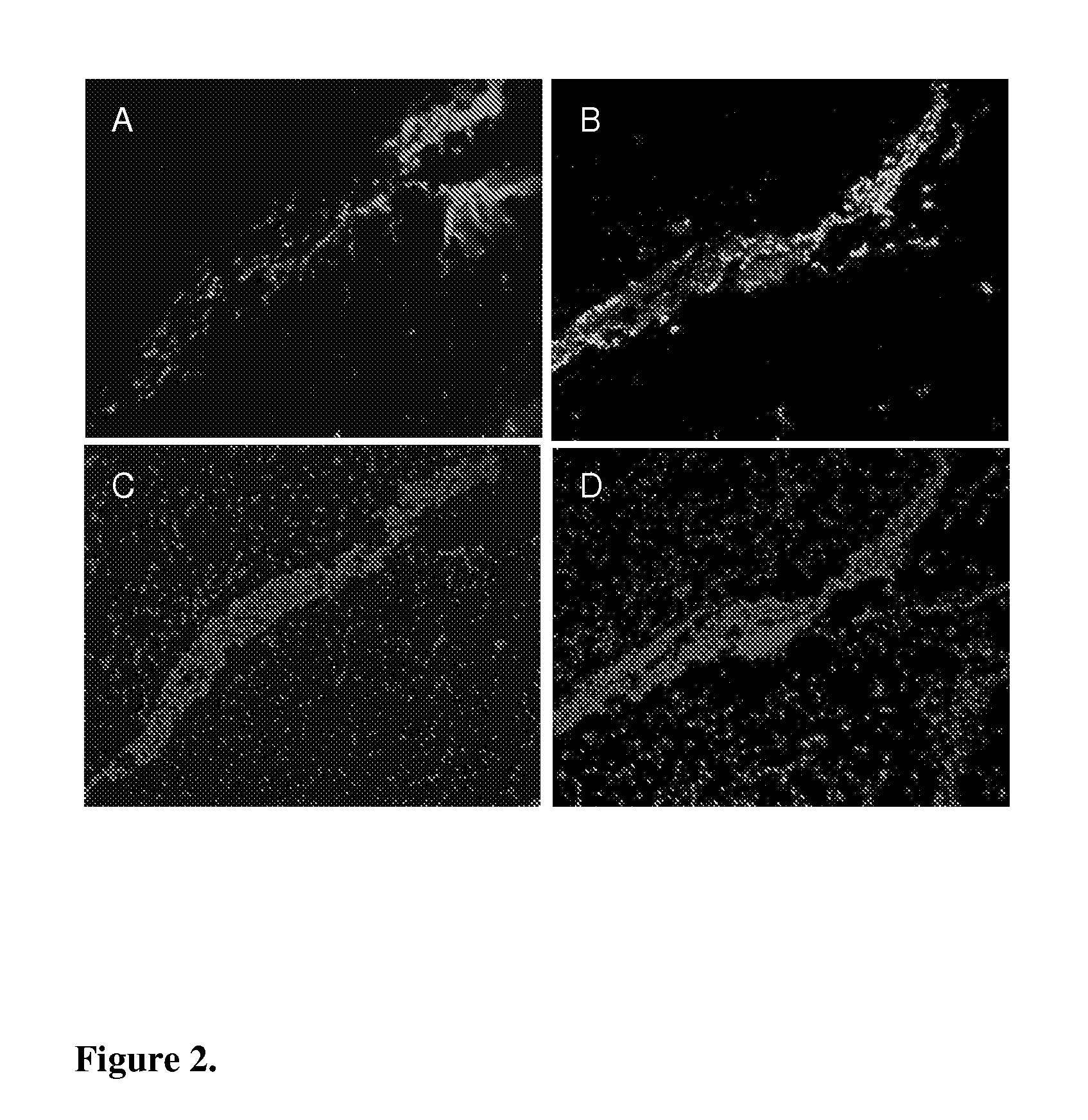
[0033]Fluorescein-conjugated CGLSGLGVA peptide (10 μg) was injected into tail vein of HIFko tumor-bearing mice. After 60 min of circulation tumor-containing brain was excised and prepared for histological analysis. Peptide was visualized by using an antibody against fluorescein (rabbit anti-FITC, Zymed Laboratories). FIG. 2 shows that the peptide (red in FIG. 2A) was detected only in the tumor islets and not in the surrounding normal brain tissue. Presence of the tumor cells was confirmed by staining for the SV40 T-antigen specific for tumor cells from a subsequent section (green in FIG. 2B). FIGS. 2C and 2D show the same microscopic field as 2A and 2B. Nuclei were visualized by DAPI staining (blue).

example 3

Fluorescein-Conjugated CGLSGLGVA Peptide Does Not Home to the Intracranial VEGF-Overexpressing Tumors

[0034]Fluorescein-conjugated CGLSGLGVA peptide (10 μg) was injected into the tail vein of VEGF overexpressing (VEGF+) tumor-bearing mice. After 60 min of circulation tumor was excised and prepared for histological analysis. Peptide was visualized by using an antibody against fluorescein (rabbit anti-FITC, Zymed Laboratories). FIG. 3A. Peptide was not detected in the tumor tissue. 3B: Same microscopic field as in 3A. Nuclei were visualized by DAPI staining (blue).


PUM

PropertyMeasurementUnit
imagingaaaaaaaaaa
distancesaaaaaaaaaa
sizeaaaaaaaaaa

Abstract

The present invention relates to a peptide, which specifically homes to the intracranial, early stage astrocytoma model that grows as islets and harbors co-opted tumor vessels in the brain. The peptide finds its use in targeted delivery of therapeutic substances to invasive brain cancer or metastatic brain lesions as such and in combination with conventional therapies, such as surgery and radiation, and anti-angiogenic therapies, and as a tool in diagnosis of, e.g., invasive brain cancer or metastatic brain lesions.

Description

FIELD OF THE INVENTION[0001]The present invention relates to a peptide, which specifically homes to the intracranial, early stage astrocytoma model that grows as islets and harbors co-opted tumor vessels in the brain, and to different glioma models. The peptide is useful in targeted delivery of therapeutic substances to brain tumors as such and in combination with conventional therapies, such as surgery and radiation, and anti-angiogenic therapies. This peptide may also be used as an effective tool in diagnosis of, e.g., invasive brain cancers and metastatic lesions of other tumor types in the brain.BACKGROUND OF THE INVENTION[0002]Astrocytes arise from multipotent neural stem cells and retain their capacity for division throughout their life span. This property is likely to make them susceptible for transformation and to contribute to the fact that astrocyte-derived tumors are the most common brain tumors in adults. Low-grade astrocytomas acquire their blood supply by propagating a...

Claims


Application Information

Patent Timeline
Patent Type & Authority Applications(United States)
IPC IPC(8): A61K38/08C07K7/06G01N33/574A61P35/00
CPCA61K38/10G01N33/57407C07K7/06A61P35/00A61P35/04A61K38/08G01N33/574G01N33/6893
Inventor LAAKKONEN, PIRJORUOSLAHTI, ERKKIBERGERS, GABRIELE
Owner BURNHAM INST FOR MEDICAL RES
Learn More