
Medical device with antimicrobial layer

a medical device and antimicrobial technology, applied in the field of medical devices, can solve the problems of prolonging hospitalization, pneumonia in ventilators, affecting the survival rate of patients, so as to promote the development of more disease resistant bacteriotypes and mitigate the colonization of the tube surfa

Inactive Publication Date: 2011-03-24
TYCO HEALTHCARE GRP LP
View PDF107 Cites 12 Cited by
  • Summary
  • Abstract
  • Description
  • Claims
  • Application Information

AI Technical Summary

Benefits of technology

The patent describes a medical device with a conduit for fluid that has a wall made of a hydrophobic polymer with an outer layer made of a hydrophilic polymer with an antimicrobial compound dispersed in it. The antimicrobial compound is made of phosphorus-based glass with a metal dispersed in it. The device is made by extruding the hydrophobic polymer to form the wall and then mixing it with the hydrophilic polymer and antimicrobial compound. The resulting device has improved antimicrobial properties and can be used for medical purposes.

Problems solved by technology

Ventilator-associated pneumonia may be a cause of morbidity in critically ill patients.
Prolongation of hospitalization, ventilation, and management of VAP infections may add up to seven days in additional patient care and over $5,000 in incremental treatment costs per patient.
Bacteria for colonization may also result from the formation of microbial adhesions or biolilms on the surfaces of contaminated medical devices.
Colonization, in turn, may lead to microaspiration of pulmonary pathogens and related lung infection.
Such interluminal biofilms may occlude the breathing tube or migrate back into the lungs to cause further infection.
The process of removing these biofilms and secretions with conventional suction catheters may lead to the aspiration of fragments of biofilms or infected aerosols.
Contaminated suction catheters, feeding tubes, ventilator tubing and breathing circuits, or filters, heat and moisture exchangers, nebulizers, heated humidifiers, or other related breathing tubes or devices may be sources of microorganism contamination and thus may contribute to biofilm formation.
Administering large doses of antibiotics, however, may promote the development of more disease resistant bacteriotypes and is thus undesirable.

Method used


Image

  • Medical device with antimicrobial layer
  • Medical device with antimicrobial layer
  • Medical device with antimicrobial layer

Examples


third embodiment

[0047]In one embodiment, the AM layer may be within a range of about 0.002 mm-2.5 mm in thickness, or about 0.13 mm in thickness. In another embodiment, the AM layer may be within a range of about 0.002 mm-2.5 mm in thickness. In a third embodiment, the AM layer may be up to about 6.35 mm in thickness. In one embodiment, substantially similar materials may form both the inner and outer surfaces of the tube.

[0048]In one embodiment, an inner or an outer AM layer may be simultaneously extruded with the medical device wall in a process commonly known as “co-extrusion.” In another embodiment, both an inner and an outer AM layer maybe extruded simultaneously with the medical device wall in a process sometimes referred to as “tri-extrusion.”

[0049]Applying an AM layer to the surface of a medical device may reduce the incidence of VAP. There may also be a production cost savings to be gained by extruding an AM layer on a medical device over a conventional coating process.

[0050]In one embodim...

first embodiment

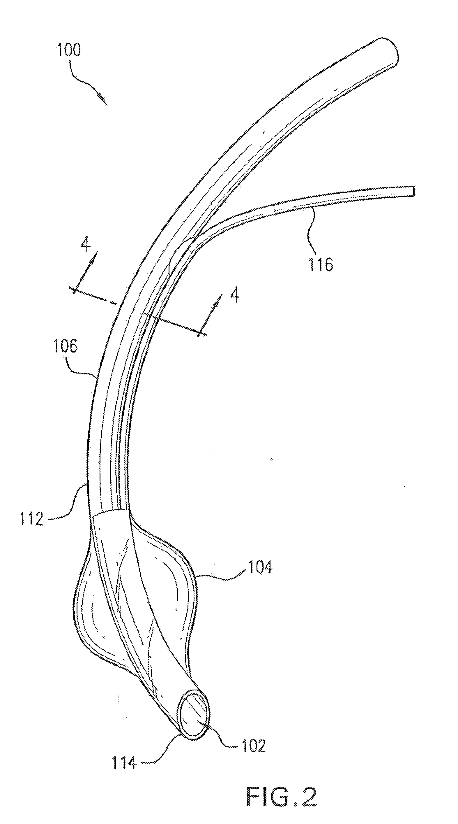
[0052]In FIG. 2 is shown a medical device 100 according to the invention. Medical device 100 may be a catheter, a stent, a feeding tube, an intravenous tube, an ET tube, a circuit, an airway accessory, a connector, an adapter, a filter, a humidifier, a nebulizer, or a prosthetic, in various embodiments.

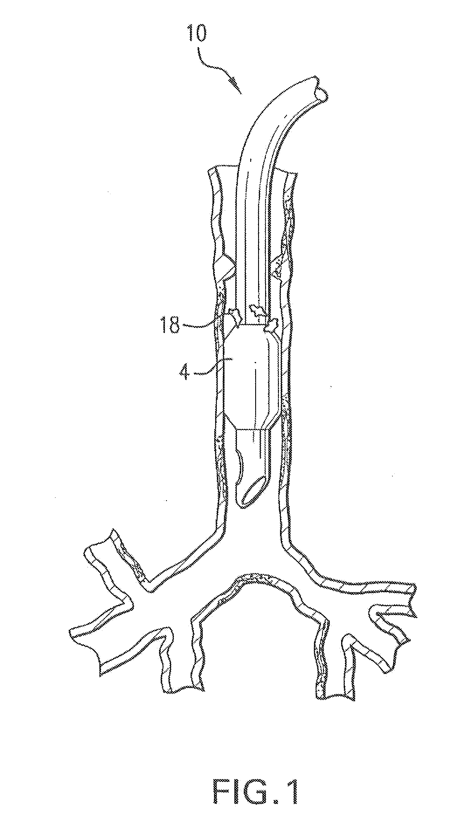
[0053]Medical device 100 may have a conduit 102 for a fluid and an inflatable cuff 104 disposed at a first end 114 of conduit 102. The fluid may be a gas, an aerosol, a suspension, a vapor, or droplets of liquid dispersed in a gas. A lumen 116 may be disposed alongside conduit 102 to inflate cuff 104. In one embodiment, a wall 112 of conduit 102 is made of a hydrophobic polymer, a hydrophilic polymer and an antimicrobial compound.

[0054]As shown in section 4-4 shown in FIG. 4, a wall 412 of conduit 102 is made of a hydrophobic polymer with an outer layer 406 composed of a hydrophilic polymer and an antimicrobial compound disposed on an outer surface 408 of wall 412. An inner layer 404 ...

second embodiment

[0056]In a second embodiment, a method of making a medical device 100 comprises the actions of providing a hydrophobic polymer, a hydrophilic polymer and an antimicrobial compound, combining the hydrophilic polymer and the antimicrobial compound, forming the hydrophobic polymer into a wall 412 of a conduit 102, and substantially simultaneously extruding the hydrophilic polymer and the antimicrobial compound as an outer layer 406 on a outer surface 408 of conduit 102.

[0057]In another embodiment, the method Further includes Forming the hydrophobic polymer into a cuff 104 on an end of conduit 102, and substantially simultaneously extruding the hydrophilic polymer and the antimicrobial compound on a surface of cuff 104.

[0058]In another embodiment, the method Further includes substantially simultaneously extruding the hydrophilic polymer and the antimicrobial compound as an inner layer 404 on an inner surface 410 of conduit 102 while wall 412 and outer layer 406 are being extruded.

[0059]...


PUM

PropertyMeasurementUnit
thicknessaaaaaaaaaa
thicknessaaaaaaaaaa
hydrophobicaaaaaaaaaa

Abstract

A medical device includes a conduit for a fluid. The conduit has a wall formed of a hydrophobic polymer with a hydrophilic polymer layer extruded over it, and an antimicrobial substantially dispersed within the hydrophilic polymer. The antimicrobial compound may be a predetermined amount of phosphorus-based glass having a predetermined quantity of a metal such as silver substantially dispersed therein. The medical device may be an endotracheal tube made by providing a hydrophobic polymer, a hydrophilic polymer and an antimicrobial compound, forming the hydrophobic polymer, the hydrophilic polymer and the antimicrobial compound into a conduit, and forming a cuff on an end of the conduit.

Description

CROSS REFERENCE TO RELATED APPLICATIONS[0001]This application is a continuation of prior U.S. application Ser. No. 11 / 337,995, filed Jan. 24, 2006, which is a continuation of prior U.S. application Ser. No. 10 / 425,030. filed Apr. 29, 2003, the specifications of which are incorporated by reference in their entirety.BACKGROUND OF THE INVENTION[0002]1. Field of the Invention[0003]The present invention relates to medical devices, and more particularly, to a method of adding an antimicrobial function to a medical device, and a system and apparatus thereof.[0004]2. Description of the Related Art[0005]Ventilator-associated pneumonia may be a cause of morbidity in critically ill patients. Approximately 250,000 cases of Ventilator Associated Pneumonia (VAP) are reported each year. The mortality associated with VAP is approximately 23,000 patients annually. (Engelmann, J. et al.; Ventilator-Associated Pneumonia; Seminars in Infection Control. Vol. 1. No. 2 2001).[0006]Prolongation of hospital...

Claims


Application Information

Patent Timeline
Patent Type & Authority Applications(United States)
IPC IPC(8): A61L31/16B32B1/08A61F2/04A61L29/16A61M16/04A61M39/00A61M39/16
CPCA61F2002/046A61L29/16A61L31/16A61L2300/102A61M16/0443A61L2300/404A61M16/04A61M39/162A61L2300/104
Inventor MARTENS, PAUL W.NIETO, ROBERT L.VIRAG, ROBERTPOTTER, ADIN M.
Owner TYCO HEALTHCARE GRP LP
Learn More