
System and method for segmenting m-mode ultrasound images showing blood vessel wall motion over time

a technology of m-mode ultrasound and motion over time, applied in the field of medical imaging, can solve the problems of limited, difficult to achieve, and difficult to segment the intima-lumen boundary, and achieve the effect of improving the quality of li

Active Publication Date: 2011-02-24
MERCK SHARP & DOHME LLC
View PDF3 Cites 22 Cited by
  • Summary
  • Abstract
  • Description
  • Claims
  • Application Information

AI Technical Summary

Benefits of technology

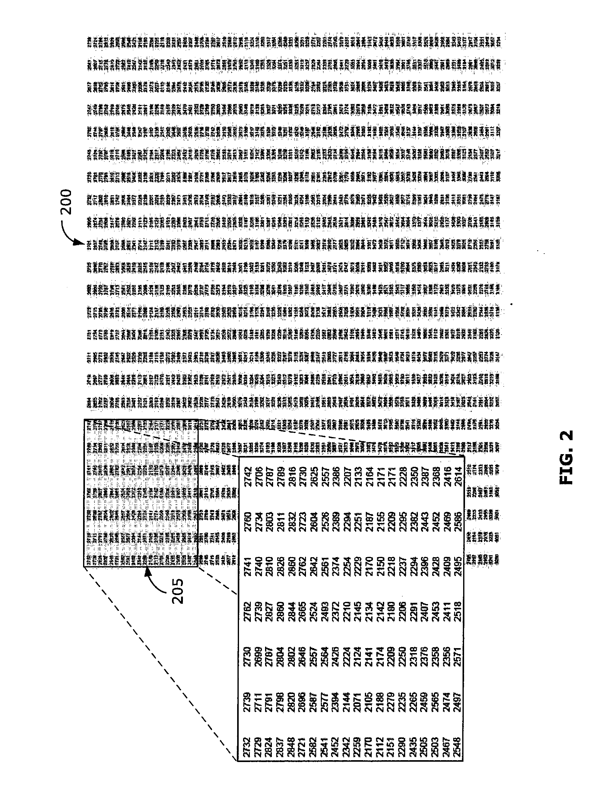
[0012]In general, systems and methods according to the present invention receive an m-mode ultrasound data file created by an ultrasound machine, store the m-mode data file on a computer-readable memory storage device and parse the data file according to a predefined m-mode data file format specification in order to produce a two-dimensional (2-D) m-mode matrix. A cross-correlation function is then applied to all pairs of scans in the 2-D m-mode matrix in order to calculate and produce a relative wall motion waveform. The cross-correlation function, which naturally exploits the banding structure typically characterizing such m-mode images, includes a global optimization procedure that enables higher resolution of the relative wall motion waveform. Then an absolute offset for the relative wall motion is calculated by interpolating the relative wall motion waveform over the 2-D m-mode matrix, and comparing the interpolated relative wall motion waveform to an established noise threshold. Ultimately, the system and method achieves segmentation of m-mode ultrasound images of blood vessels to sub-pixel resolution. Since the process is completely automated, it is more reliable than existing manual techniques.
[0027](G) using the segmentation program to cause the microprocessor to calculate and store on the memory storage device an absolute offset based on the relative wall motion waveform.

Problems solved by technology

Because only a single direction is probed, the PRF can be very fast, limited only by the time required for the pulse to reach the depth of interest and return.
For a number of technical reasons, accurately and reliably identifying, mapping and displaying the absolute waveform corresponding to the location and movement of the intima-lumen boundary in an m-mode ultrasound image of a blood vessel remains a significant and difficult challenge using conventional systems and techniques.
Various systems and techniques for segmenting the intima-lumen boundary have been proposed and used with limited success.
However, by using only local information, the method is not robust in extended regions of weak returns.
However, this method typically results in a reduced, and therefore misleading, estimate of wall distension.

Method used


Image

  • System and method for segmenting m-mode ultrasound images showing blood vessel wall motion over time
  • System and method for segmenting m-mode ultrasound images showing blood vessel wall motion over time
  • System and method for segmenting m-mode ultrasound images showing blood vessel wall motion over time

Examples


Embodiment Construction

[0053]Exemplary embodiments of the invention will now be described in more detail with reference to the figures. First, there is provided a detailed discussion of the computational elements and mathematical equations encompassed by computer programs (e.g., software) adapted to operate according to exemplary embodiments of the invention, followed by a detailed description of some of the hardware components and program flows that may be incorporated into such exemplary embodiments.

Computational Elements and Mathematical Equations Encompassed by the Segmentation Program

[0054]The computer system of the present invention is preprogrammed with a plurality of program modules (collectively referred to herein as the “segmentation program”) having computer-readable instructions adapted to cause the microprocessor to apply a series of mathematical calculations to the pixel intensity data in the scans of an m-mode data matrix, as described below, to achieve the practical result of producing bot...


PUM


Abstract

The present invention uses a microprocessor, a memory storage device, and a segmentation program comprising a plurality of program modules containing computer-readable instructions that cause the microprocessor to measure the spatial offsets between all pairs of scans in an m-mode image of a blood vessel with a cross-correlation function, convert the spatial offsets to a relative wall motion waveform through a global optimization procedure, and then translate the relative wall motion waveform to an absolute wall motion waveform by interpolation over the m-mode image. The resulting detailed absolute wall distension waveform may be beneficially rendered (e.g., superimposed) on the m-mode ultrasound image for display (e.g., on a printer and / or video monitor) and diagnostic purposes, and has enormous potential for enhancing existing techniques for identifying and studying vascular biomarkers, such as vessel wall strain and compliance.

Description

CROSS-REFERENCE TO RELATED APPLICATIONS[0001]Not Applicable.FIELD OF THE INVENTION[0002]The present invention pertains to medical imaging. More particularly, it relates to systems and methods for analyzing and segmenting m-mode ultrasound images showing the movement of blood vessel walls over time.RELATED ART[0003]The mechanical state of a blood vessel at a given point and time can be well described using wall area, blood pressure and flow, and their control. A large artery wall, for example, is composed of three layers, namely the tunica intima, tunica media and tunica adventitia, each playing different roles in the function and dynamic response of the vessel wall to stresses. The artery wall is compliant and stretches in response to pressure. The average artery wall diameter may also change in response to its environment, a process known as remodeling. Both the compliance and the remodeling aspects of the blood vessel wall can change in short term regulation, long term regulation,...

Claims


Application Information

Patent Timeline
IPC IPC(8): G06K9/00
CPCG06K2209/05G06T2207/30104G06T2207/10132G06T7/2033G06T7/246G06V2201/03
Inventor FANCOURT, CRAIGAZER, KARIMWALKER, III, MATTHEW
Owner MERCK SHARP & DOHME LLC
Learn More