
Medical image interpretation system

a medical image and image technology, applied in tomography, applications, instruments, etc., can solve the problems of time-consuming, laborious, and inability to selectively select the segmentation technique,

Active Publication Date: 2011-01-06
TOSHIBA MEDICAL SYST CORP
View PDF14 Cites 31 Cited by
  • Summary
  • Abstract
  • Description
  • Claims
  • Application Information

AI Technical Summary

Problems solved by technology

However, the segmentation technique is not for selectively specifying a region described in a finding or diagnosis of a diagnostic report.
It takes time to perform this operation.
In particular, in a case that a physician who is not engaged in interpretation refers to a diagnostic report, it takes effort to search out a finding provided with no specified key image from among images.

Method used


Image

  • Medical image interpretation system
  • Medical image interpretation system
  • Medical image interpretation system

Examples


first embodiment

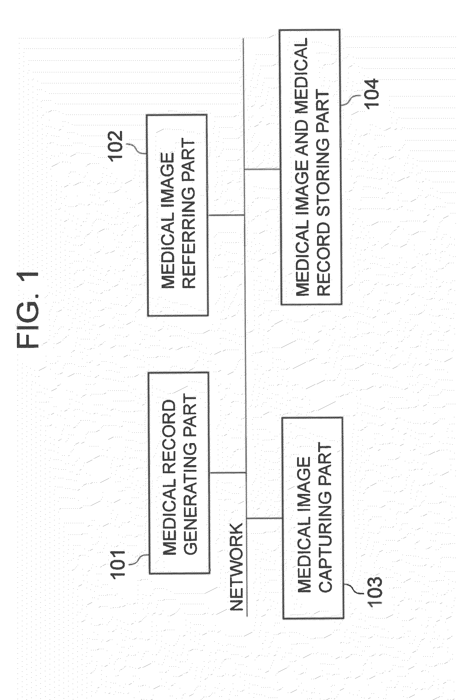
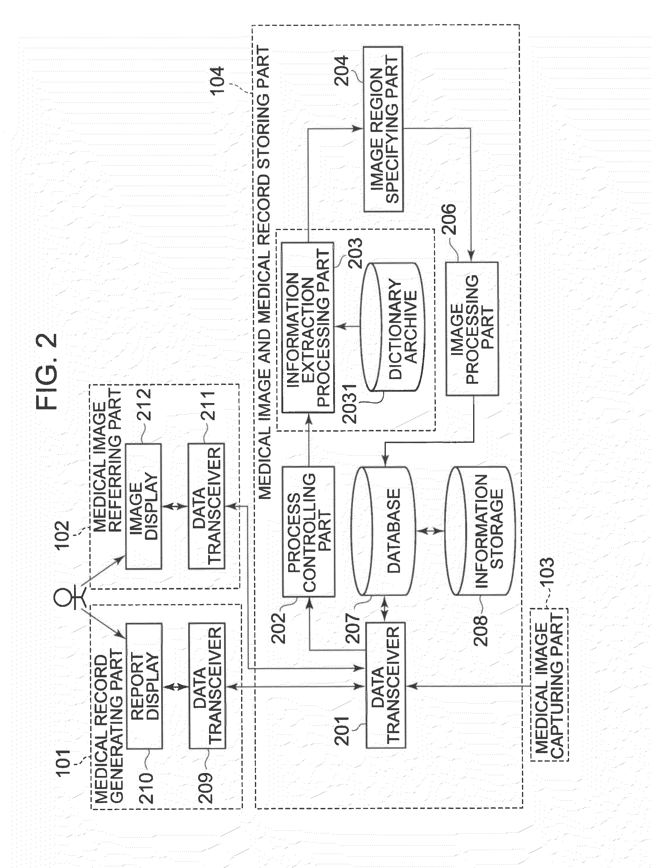
[0033]Below, a medical image interpretation system relating to a first embodiment will be described with reference to FIGS. 1 and 2.

[0034]Hereinafter, a “medical record” refers to data including text data of a finding or the like associated with a medical image, such as a diagnostic report and a medical chart. A “medical image” refers to an image of a patient captured with a medical imaging device, and may be composed of a plurality of images as an image captured with a CT or an MRI is, or may be composed of one image as an X-ray image is.

[0035]Hereinafter, a case will be explained in which the medical image is composed of a plurality of images. Below, a case of processing a diagnostic report as the medical record will be described as an example.

[0036]The medical image interpretation system relating to this embodiment specifies a region for which a finding or a diagnosis is written on a previous diagnosis report of a patient (hereinafter, the “finding” shall include the content of a...

second embodiment

[0109]Next, a medical image interpretation system relating to a second embodiment will be described with reference to FIG. 10. FIG. 10 is a system block diagram of the medical image interpretation system relating to this embodiment. Below, a case of processing a diagnostic report as a medical record will be described as an example.

[0110]In the medical image interpretation system relating to this embodiment, while the operator (the radiologist) is generating a diagnostic report on a present examination image with the medical record generating part 101, the medical image interpretation system relating to this embodiment, at a point that a sentence being described is fixed, structures the sentence and extracts a region term, specifies the position and range of a region, and links images showing the region to the sentence or pastes the images to the diagnostic report.

[Configuration]

[0111]Firstly, components that configure the medical image interpretation system relating to this embodime...


PUM


Abstract

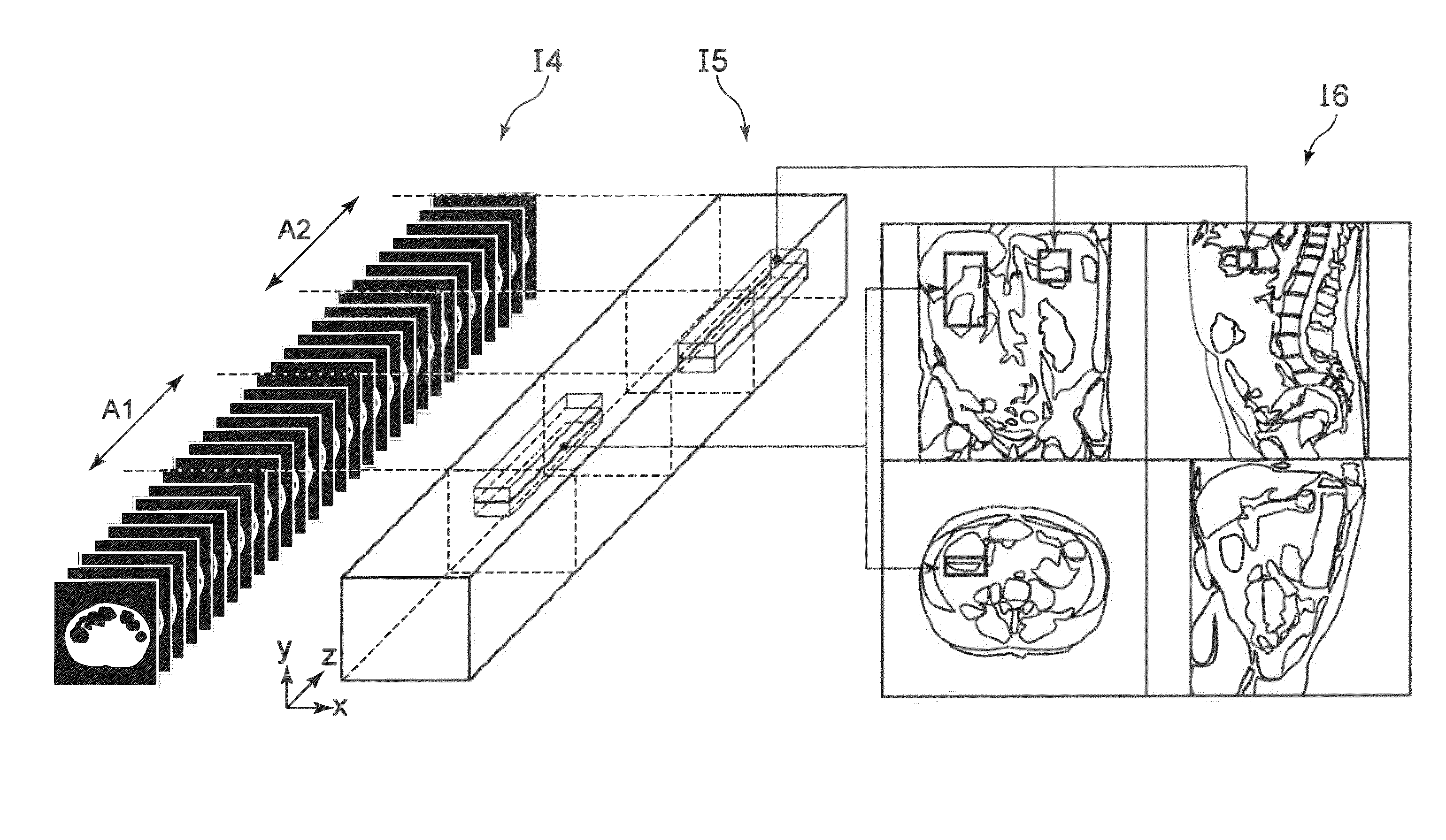
In embodiments, a medical image interpretation system has at least a medical image storing part, a medical image referring part, a medical record storing part, a process controlling part, an information extraction processing part, an image region specifying part, and an image processing part. The process controlling part specifies a medical image and extracts a medical record of a patient of the medical image. The information extraction processing part classifies terms on a sentence written in the medical record into predetermined types to structure the sentence, and extracts a region term from the structured sentence. The image region specifying part specifies the range of images showing a region corresponding to the region term from the medical image and specifies the position and range of the region. The image processing part displays at least the region position and range in a visually recognizable manner.

Description

CROSS-REFERENCE TO RELATED APPLICATION[0001]This application is based upon and claims the benefit of priority from Japanese Patent Application No. 2009-157979, filed on Jul. 2, 2009; the entire contents of which are incorporated herein by reference.BACKGROUND[0002]1. Field[0003]Embodiments described herein relate generally to a medical image interpretation system in which a medical image storing apparatus that stores images of patients captured with a medical imaging device such as an X-ray computed tomography (CT) apparatus or a magnetic resonance imaging (MRI) apparatus, an image referring apparatus that allows a user to refer to the stored images, a medical record generating apparatus that generates medical finding reports on images, and a medical record referring apparatus that refers to the generated medical records are connected via a network.[0004]2. Description of the Related Art[0005]Conventionally, such a medical image interpretation system has been utilized that a medical...

Claims


Application Information

Patent Timeline
IPC IPC(8): G06K9/00A61B5/00A61B5/055A61B6/03G06F17/30G06T1/00G16H10/60G16H15/00G16H30/40
CPCG06F19/321G06T7/0012G06F19/3487G06F19/322G16H10/60G16H15/00G16H30/40
Inventor FUTAMI, HIKARUYAMAGISHI, HIROMASA
Owner TOSHIBA MEDICAL SYST CORP
Learn More