
Segmentation of regions in measurements of a body based on a deformable model

a segmentation and model technology, applied in the field of spatial segmentation measured values of a body, can solve the problems of inability to provide a sharp contrast edge in the measured value, the segmentation process to determine the significant regions in the measured value set is tedious and time-consuming, and the segmentation process cannot handle large data sets. to achieve the effect of reducing the measurement of curvatur

Inactive Publication Date: 2010-02-04
THE CLEVELAND CLINIC FOUND +1
View PDF5 Cites 88 Cited by
  • Summary
  • Abstract
  • Description
  • Claims
  • Application Information

AI Technical Summary

Benefits of technology

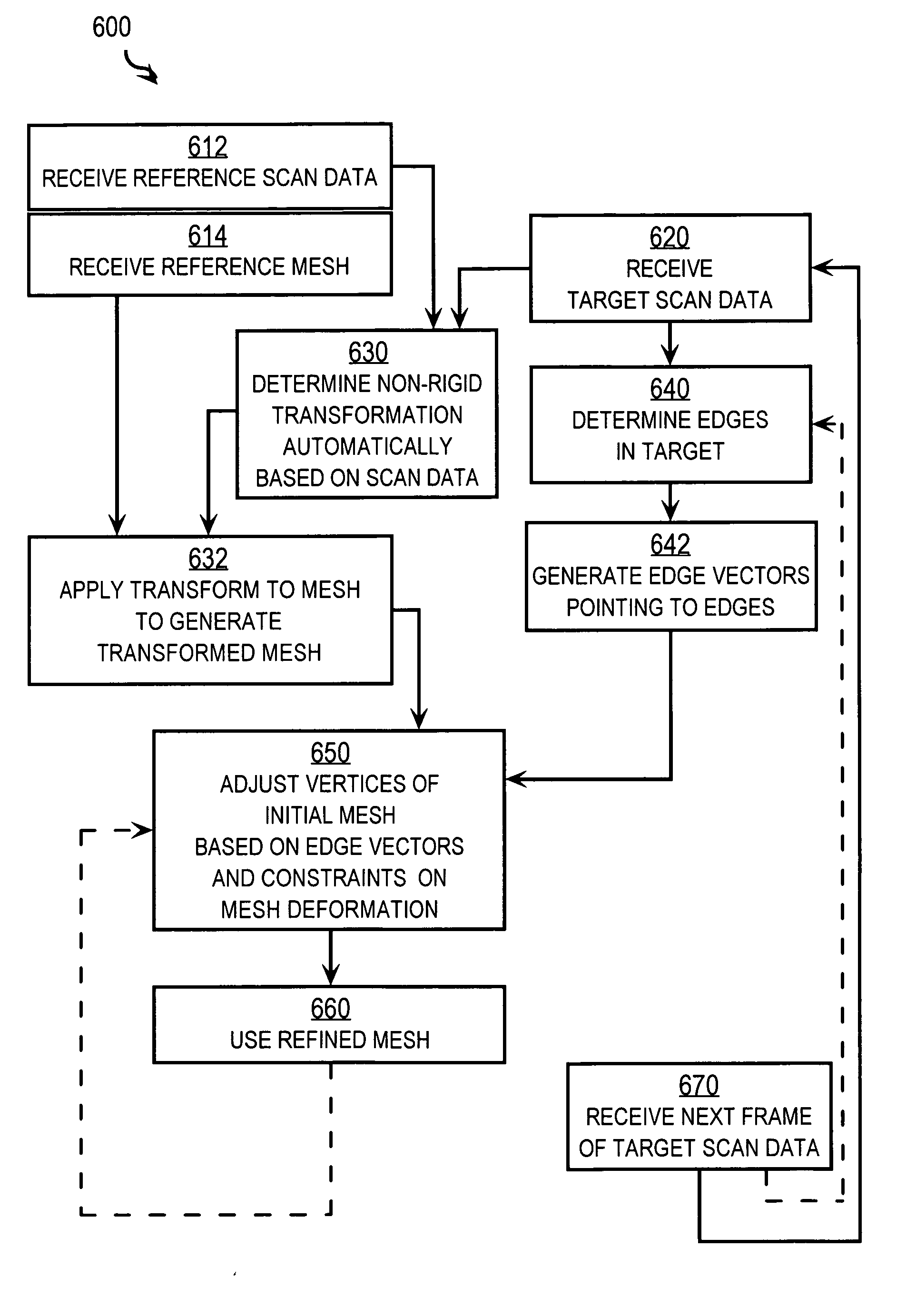
[0014]In another set of embodiments, a method includes receiving reference segmentation data and target data. The reference segmentation data includes coordinate values for each vertex in a first set of vertices that indicates a first boundary of a myocardial wall of a body, and coordinate values for each vertex in a second set of vertices that indicates a second boundary of the myocardial wall. The target data includes values based on a measurement mode of measuring the body. Each target data value is associated with coordinate values. A coordinate for a first vertex of the first set is adjusted toward a higher gradient in values of the target data in a vicinity of the first vertex, by determining a local separation distance from the first vertex to the second boundary, and adjusting the coordinate based on the local separation distance.
[0015]In another set of embodiments, a method includes receiving reference segmentation data and target data. The reference segmentation data includes coordinate values for spatial or temporal dimensions for each vertex in a set of vertices that indicates a boundary of a physical component of a body. The target data includes values based on a measurement mode of measuring the body. Each target data value is associated with coordinate values. At least one coordinate for at least one vertex of the set of vertices is adjusted toward a higher gradient in values of the target data in a vicinity of the vertex by computing a measure of curvature at the vertex. It is determined whether the measure of curvature exceeds a threshold curvature. If so, then at least one coordinate of the vertex is changed to reduce the measure of curvature. As a result after all adjustments in an absence of a higher gradient, the plurality of vertices form a non-circular shape in at least one plane.
[0016]In another set of embodiments, a method includes receiving segmentation data and target data. The segmentation data includes coordinate values for spatial or temporal dimensions for each vertex in a set of vertices that indicates a boundary of a physical component of a body. The target data includes values based on a m

Problems solved by technology

However, many systems give relative intensities of measured values within the target body but do not automatically identify a particular intensity boundary with the edge of a particular structural component.
For example, some human tissues of different organs have similar intensities and do not provide a sharp contrast edge in the measured values.
In general, the segmentation process to determine the significant regions in a set of measured values is tedious and time consuming with substantial input from a human expert.
While suitable in many circumstances, there are deficiencies for some circumstances.
One deficiency is that such segmentation can not handle large data sets.
Manual segmentation for large data sets, such as those produced by real-time 3D echocardiography, remains excessively cumbersome, and thus unsuitable for routine clinical use.
Another deficiency is that segmentation with manual steps can not keep pace with body operations.
The movement can be monitored with near real time CT scans or real time ultrasound scans, but the resulting measurements would not be segmented in a timely fashion using manual approaches, and the tumor may be hard to identify and treat effectively without collateral damage to healthy surrounding tissue.

Method used


Image

  • Segmentation of regions in measurements of a body based on a deformable model
  • Segmentation of regions in measurements of a body based on a deformable model
  • Segmentation of regions in measurements of a body based on a deformable model

Examples


Embodiment Construction

[0002]This invention was made with Government support under Grant No. DAMD17-99-1-9034 awarded by the Department of Defense and Grant No. RG01-0071 awarded by the Whitaker Foundation. The Government has certain rights in the invention.

BACKGROUND OF THE INVENTION

[0003]1. Field of the Invention

[0004]The present invention relates to spatially segmenting measured values of a body, such as a patient, to interpret structural or functional components of the body; and, in particular to automatically segmenting three dimensional scan data such as Computer Tomography X-Ray (CT) scan data and echogram data.

[0005]2. Description of the Related Art

[0006]Different sensing systems are widely known and used for non-invasively probing the interior structure of bodies. For example, X-rays and X-ray-based computer-aided tomography (CT), nuclear magnetic resonance (NMR) and NMR-based magnetic resonance imagery (MRI), acoustic waves and acoustics-based ultrasound imagery (USI), positron emissions and pos...


PUM


Abstract

Techniques for segmenting data include receiving reference segmentation data and target data. The reference segmentation data defines a mesh indicating a boundary of a physical component in a reference body. The target data includes measured values at coordinates within a target body. Coordinates for vertices of the mesh are moved toward nearby edges in values of the target data. The adjustment also may be based on deviations from adjacent vertices or from a three dimensional generalized gradient vector field. The mesh may be initially transformed by a particular transformation that automatically maximizes a similarity measure between the target data and reference data. The reference data includes measured values within the reference body. The reference segmentation also may define a second mesh that indicates a second boundary in the reference body, and the adjustment is also based on an adjusted distance between the mesh and the second mesh.

Description

CROSS-REFERENCE TO RELATED APPLICATIONS[0001]This application claims benefit of Provisional Appln. 60 / 712,629, filed Aug. 30, 2005, the entire contents of which are hereby incorporated by reference as if fully set forth herein, under 35 U.S.C. §119(e).STATEMENT OF GOVERNMENTAL INTEREST[0002]This invention was made with Government support under Grant No. DAMD17-99-1-9034 awarded by the Department of Defense and Grant No. RG01-0071 awarded by the Whitaker Foundation. The Government has certain rights in the invention.BACKGROUND OF THE INVENTION[0003]1. Field of the Invention[0004]The present invention relates to spatially segmenting measured values of a body, such as a patient, to interpret structural or functional components of the body; and, in particular to automatically segmenting three dimensional scan data such as Computer Tomography X-Ray (CT) scan data and echogram data.[0005]2. Description of the Related Art[0006]Different sensing systems are widely known and used for non-inv...

Claims


Application Information

Patent Timeline
IPC IPC(8): G06K9/00
CPCG06K9/6207G06T7/0089G06T7/0083G06T7/12G06T7/149G06V10/755
Inventor SHEKHAR, RAJZAGRODSKY, VLADIMIRWALIMBE, VIVEK
Owner THE CLEVELAND CLINIC FOUND
Learn More