
Method And Composition For Proctecting Neuronal Tissue From Damage Induced By Elevated Glutamate Levels

a technology of glutamate and neuronal tissue, which is applied in the direction of drug compositions, biocides, peptide/protein ingredients, etc., can solve the problems of neuronal death, excessive stimulation, and accelerated depletion of these limited energy resources, so as to reduce blood glutamate levels, and reduce extracellular brain glutamate levels

Inactive Publication Date: 2009-12-10
MOR RES APPL LTD +1
View PDF0 Cites 12 Cited by
  • Summary
  • Abstract
  • Description
  • Claims
  • Application Information

AI Technical Summary

Benefits of technology

[0021]According to one aspect of the present invention there is provided a method of reducing extracellular brain glutamate levels, the method comprising administering to a subject in need thereof an agent capable of modulating stress hormone activity thereby reducing blood glutamate levels, thereby reducing extracellular brain glutamate levels.
[0024]According to still another aspect of the present invention there is provided a method of reducing extracellular brain glutamate levels in a subject in need thereof, the method comprising: (a) obtaining a blood sample; (b) contacting the blood sample with an agent capable of modulating stress hormone activity thereby reducing glutamate levels of cells present in the blood sample to thereby obtain glutamate depleted blood cells; and (c) introducing the glutamate depleted blood cells into the subject, thereby reducing extracellular brain glutamate levels thereof.
[0062]The present invention successfully addresses the shortcomings of the presently known configurations by providing methods and compositions for protecting neuronal tissue from damage induced by elevated glutamate levels

Problems solved by technology

However, failure or reduction in the transport process such as under ischemic conditions, results in accumulation of glutamate in the extracellular synaptic fluid and excessive stimulation of excitatory receptors, a situation that leads to neuronal death.
Two additional factors complicate and make matters worse: (i) overstimulated neurons begin to release excessive quantities of glutamate at additional synaptic junctions; this causes even more neurons to become overstimulated, drawing them into a neurotoxic cascade that reaches beyond the initial zone of ischemia; and, (ii) overstimulated neurons begin utilizing any available supplies of glucose or oxygen even faster than normal, which leads to accelerated depletion of these limited energy resources and further impairment of the glutamate transport process.
This biochemical cascade of induction and progression may continue for hours or days and causes delayed neuronal death.
Though displaying powerful neuroprotective effects in experimental stroke and head trauma, the glutamate receptor antagonists failed in clinical trials mainly because of their adverse or even lethal effects (Birmingham, 2002; Lutsep and Clark, 2001; Palmer, 2001).
However, none of the above-described approaches have been successful in providing a viable therapeutic approach for lowering glutamate levels.

Method used


Image

  • Method And Composition For Proctecting Neuronal Tissue From Damage Induced By Elevated Glutamate Levels
  • Method And Composition For Proctecting Neuronal Tissue From Damage Induced By Elevated Glutamate Levels
  • Method And Composition For Proctecting Neuronal Tissue From Damage Induced By Elevated Glutamate Levels

Examples


example 1

Brain Excess Glu Levels are Regulated by a Brain-to-Blood Glu Efflux

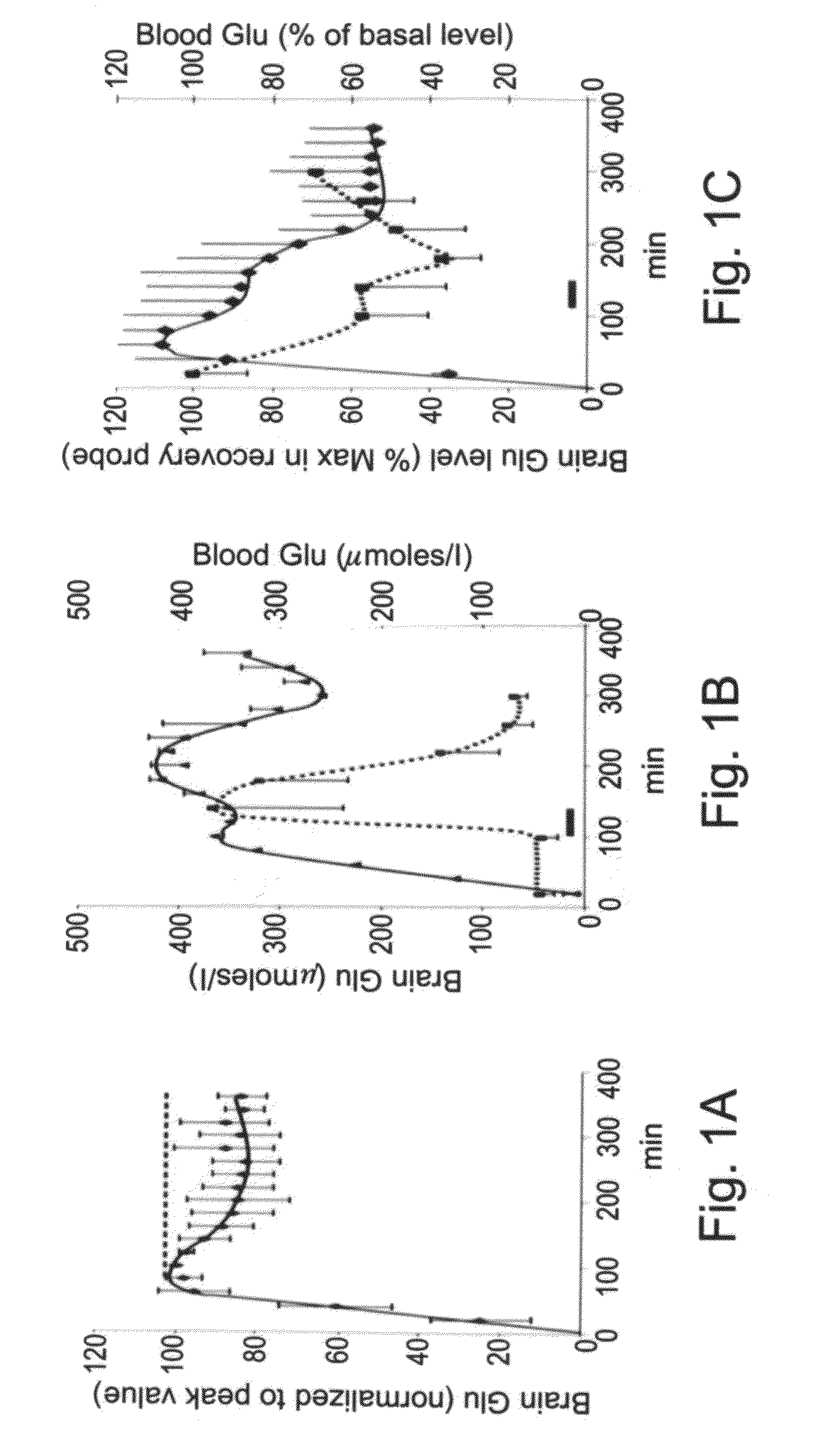
[0186]Glu efflux from the brain parenchyma interstitial fluid (ISF) to blood was first assessed [10]. The dual-probe brain microdialysis was used and perfused through the delivery probe inserted in the striatum of anesthetized rats a Glu solution while simultaneously perfusing artificial CSF through a recovery probe, located at 1 mm distance. Studies of dual-probe microdialysis describe a tissue delivery of solutes from the delivery probe of 3-6% [12] and a solute recovery of 5% in the recovery probe [13]. As Glu flows out of the first probe, it diffuses through the brain parenchyma where it is taken up into glial cells, neurons and blood capillary endothelial cells. However, as Glu keeps oozing from the delivery probe and saturates the transporters, it eventually reaches the recovery probe if its starting concentration is sufficiently high (>0.5M).

[0187]FIG. 1A shows that linearly increasing amounts of Glu arrive w...

example 2

Regulation of Blood Glu Levels by Stress

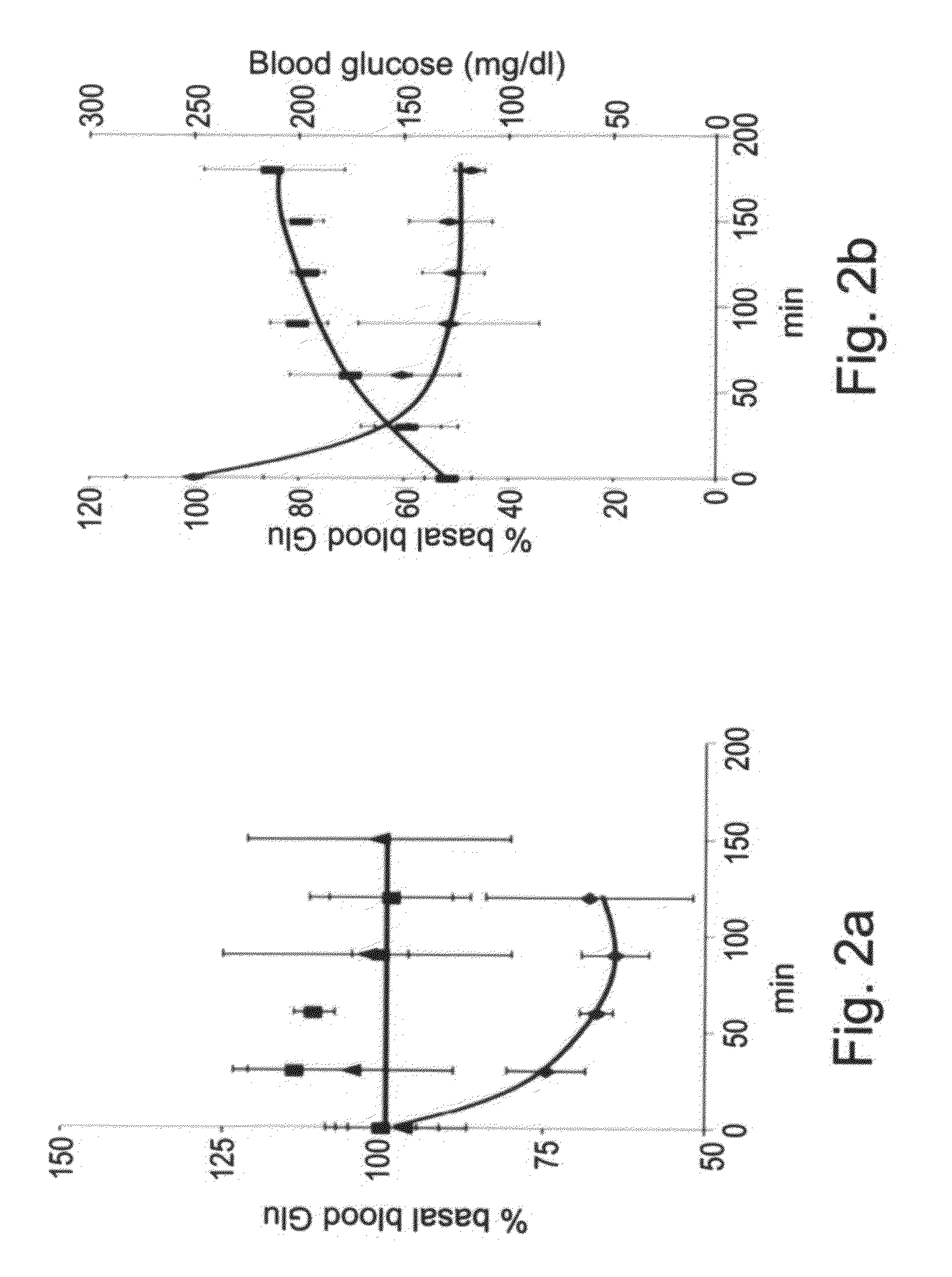
[0193]What causes the spontaneous decrease of blood Glu levels? It was suggested that such decrease could be part of a general stress response activating the hypothalamo-pituitary-adrenal (HPA) axis and the sympathetic nervous system (SNS) [15]. Since the stress response involves the release into blood of stress hormones such as cortisol, noradrenaline and adrenaline, their respective effects on the blood levels of glutamate and glucose were tested, the latter serving as a stress marker [16, 17].

[0194]FIG. 2A illustrates the fact that while neither cortisol nor noradrenaline affected blood Glu levels, adrenaline, administered over 30 min, caused a sustained blood Glu decrease to about 60% of its basal levels. As excess brain Glu could cause the activation of a stress response [15], we tested the effects of the mere insertion into brain parenchyma of a microdialysis probe. FIG. 2B shows that the probe insertion is a stressful procedure as it ca...

example 3

Stress Causes a Spontaneous Recovery from Traumatic Brain Injury

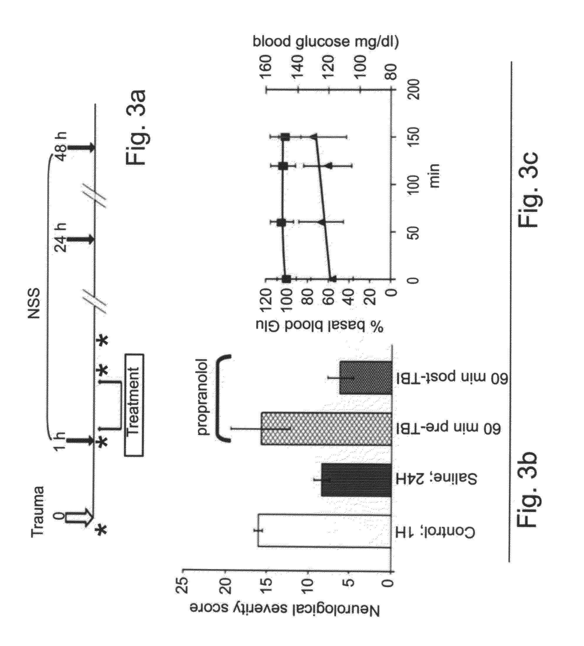
[0195]The decrease of blood Glu levels caused by stress was then addressed for its ability to neuroprotect from a brain insult well known to produce excess Glu levels in the brain of rodents [18-20] and of human victims of head injury [19, 21-23].

[0196]To this end, rats were subjected to a traumatic brain injury (TBI) [24], assessed a neurological severity score (NSS) 1, 24 and 48 h post injury, and monitored in parallel the blood Glu levels as described in FIG. 3A. The inflicted brain injury was very severe as expressed by the large NSS value of 16.2+ / −0.5 (n=33) measured at 1 h post TBI (FIG. 3C-left first column). Animals appear nevertheless to rapidly recover since, in comparison to the NSS measured at 1 h post TBI, there was a very significant NSS improvement after 24 h (FIG. 3C-first two columns). As such spontaneous improvement could possibly be attributed to the stress / adrenaline-induced decrease of blood Glu le...


PUM

PropertyMeasurementUnit
stress hormone activityaaaaaaaaaa
concentrationaaaaaaaaaa
concentrationsaaaaaaaaaa

Abstract

A method of reducing extracellular brain glutamate levels is provided. The method comprising administering to a subject in need thereof an agent capable of modulating stress hormone activity thereby reducing blood glutamate levels, thereby reducing extracellular brain glutamate levels.

Description

FIELD AND BACKGROUND OF THE INVENTION[0001]The present invention relates to a method and composition for protecting the central nervous system (CNS) from damage induced by abnormal levels of glutamate, which may result from, for example, a stroke.[0002]The central nervous system is composed of trillions of nerve cells (neurons) that form networks capable of performing exceedingly complex functions.[0003]The amino acid L-glutamic acid (Glutamate), mediates many of the excitatory transactions between neurons in the central nervous system. Under normal conditions, accumulation of glutamate in the extracellular space is prevented by the operation of a recycling mechanism that serves to maintain neuronal glutamate levels despite continual loss through transmitter release (Van der Berg and Garfinkel, 1971; Kennedy et al., 1974). Glutamate, released by glutamatergic neurons, is taken up into glial cells where it is converted into glutamine by the enzyme glutamine synthetase. Glutamine reen...

Claims


Application Information

Patent Timeline
Patent Type & Authority Applications(United States)
IPC IPC(8): A61K45/00A61K38/43A61K38/45A61K38/44A61P25/00
CPCA61K31/198A61P25/00A61P25/02A61P25/28A61P43/00
Inventor TEICHBERG, VIVIAN I.ZLOTNIK, ALEXANDERSHAPIRA, YORAM
Owner MOR RES APPL LTD
Learn More