
Composition and method for diagnosing esophageal cancer and metastasis of esophageal cancer

Inactive Publication Date: 2009-10-29
TORAY IND INC +1
View PDF2 Cites 20 Cited by
  • Summary
  • Abstract
  • Description
  • Claims
  • Application Information

AI Technical Summary

Benefits of technology

[0178]The present invention provides a composition for determining the disease, i.e. esophageal cancer or metastasis of esophageal cancer, wherein the composition is useful for diagnosing, detecting, determining, or predicting the esophageal cancer or the metastasis of esophageal cancer, and for treating the esophageal cancer. The present invention further provides a method for diagnosing, detecting, determining, or predicting the esophageal cancer and the metastasis of esophageal cancer, using said composition. Use of the composition produces a remarkable advantage that is to provide a simple, highly predictable, rapid and simple method for detecting, determining, or predicting the esophageal cancer or the metastasis of esophageal cancer.
[0179]Some of the esophageal cancer markers of the invention can be observed in biological samples, such as blood, from patients with esophageal cancer; however, they are not or almost not found in healthy persons. For this reason, using the presence or amount of such markers as an indicator enables easy detection of esophageal cancer in the blood, for example.

Problems solved by technology

Surgery, particularly extensive regional lymph node dissection, imposes serious burdens upon patients, and thus, lowered QOL after surgery should be an issue of concern.
These symptoms, however, occur as a result of the growth of cancer in the esophagus, and the cancer, which is found at the time of consultation following self-detection, has already progressed or metastasized outside the esophageal wall, and such a cancer often indicates a poor prognosis.
However, these markers have poor sensitivity and specificity, and the sensitivity of CYFRA21-1, which is likely to have the highest sensitivity, is as low as about 33.9% (Nakamura, T. et al., 1998, Diseases of the Esophagus, vol.
Thus, this technique has not yet enabled to determine the presence or absence of esophageal cancer cells by detecting said serum markers alone or in combination, nor is it possible to diagnose the metastasis of esophageal cancer using said markers.
The aforementioned known indicators, however, are disadvantageously poor in specificity and / or sensitivity.
Also, a method for effectively detecting such indicators from biological samples has not yet been established.
Additionally, at present, it is considered impossible to detect the presence or absence of the metastasis of esophageal cancer, which is a key factor in determining a patient's prognosis, using diagnostic markers.

Method used


Image

  • Composition and method for diagnosing esophageal cancer and metastasis of esophageal cancer
  • Composition and method for diagnosing esophageal cancer and metastasis of esophageal cancer
  • Composition and method for diagnosing esophageal cancer and metastasis of esophageal cancer

Examples


example 1

1. Clinical and Pathological Findings Concerning Subjects

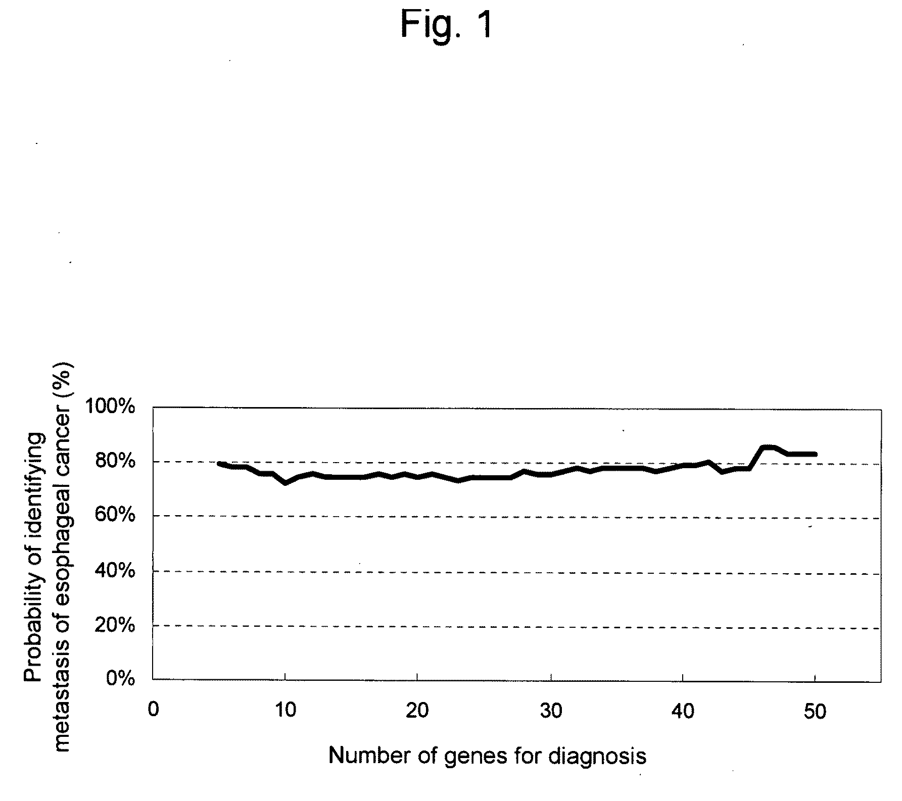
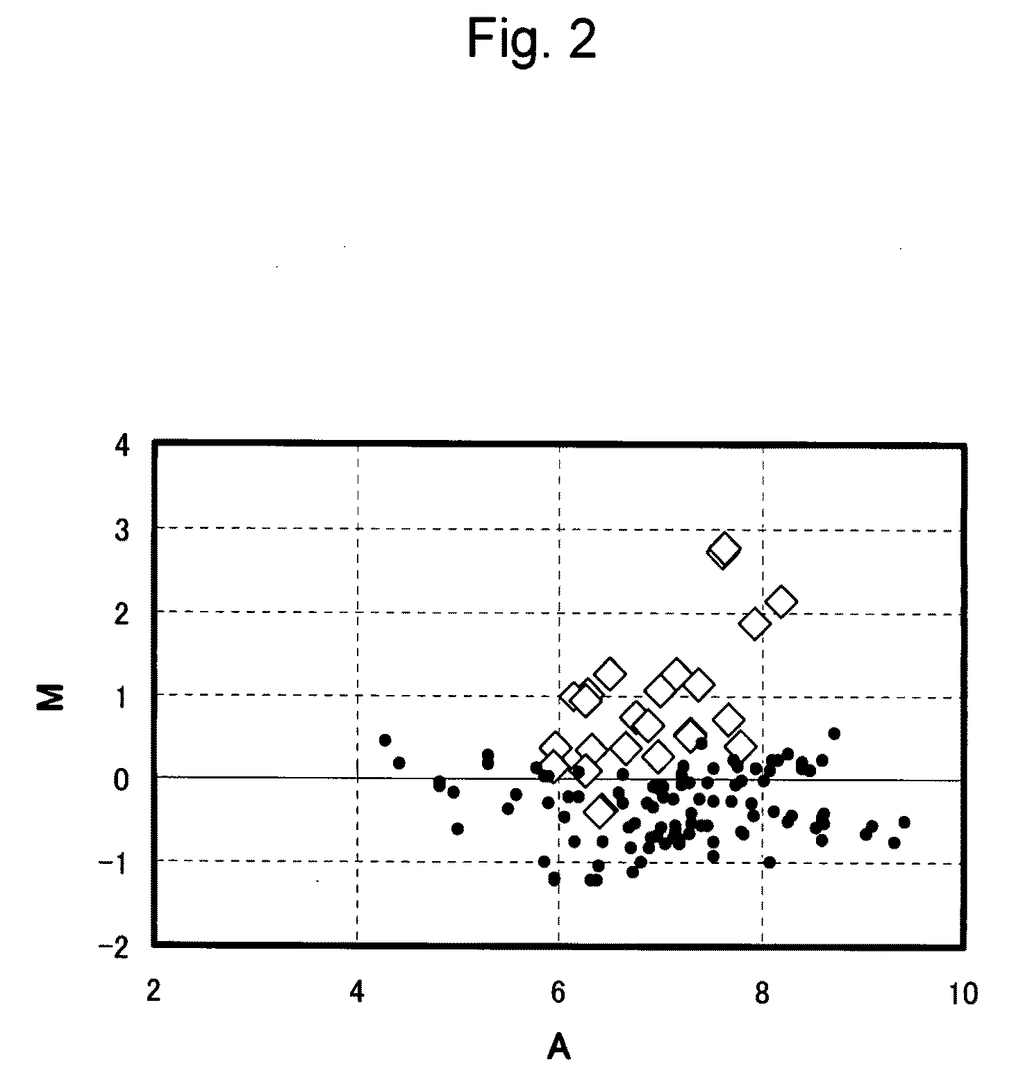
[0635]Informed consent was obtained from 119 patients with esophageal cancer, and the esophagus tissues were excised from them at the time of the surgical excision of esophageal cancer or the esophageal biopsy. Part of the excised tissue was visually and / or histopathologically inspected to identify the esophageal cancer tissue, the esophageal cancer lesions were separated from the normal tissue, both of which were immediately freezed and stored in liquid nitrogen. Separately, the regional lymph nodes in the vicinity were removed from the excised tissue in order to pathologically diagnose the presence or absence of the metastasis of esophageal cancer cells.

2. Extraction of Total RNA and Preparation of cDNA

[0636]The tissue in the esophageal cancer lesion of the esophageal tissue obtained from an esophageal cancer patient was used as a sample. Total RNA was prepared from the tissue using a Trizol reagent (Invitrogen) in accordanc...

example 2

1. Clinical and Pathological Findings Concerning Subjects

[0646]Informed consent was obtained from 119 patients with esophageal cancer, and the esophagus tissues were excised from them at the time of the surgical excision of esophageal cancer or the esophageal biopsy. Part of the excised tissue slices was visually and / or histopathologically inspected to identify the esophageal cancer tissue, the esophageal cancer lesions were separated from the normal tissue, both of which were immediately freezed and stored in liquid nitrogen.

2. Extraction of Total RNA and Preparation of cDNA

[0647]The tissue in the esophageal cancer lesion of the esophageal tissue and the non-cancerous tissue (normal tissue) in the same esophageal tissue, which tissues had been obtained from a patient with esophageal cancer, were used as samples. Total RNA was prepared from the tissues using a Trizol reagent (Invitrogen) in accordance with the manufacturer's recommended protocol.

[0648]The thus obtained total RNA (1 ...

example 3

1. Detection by RT-PCR

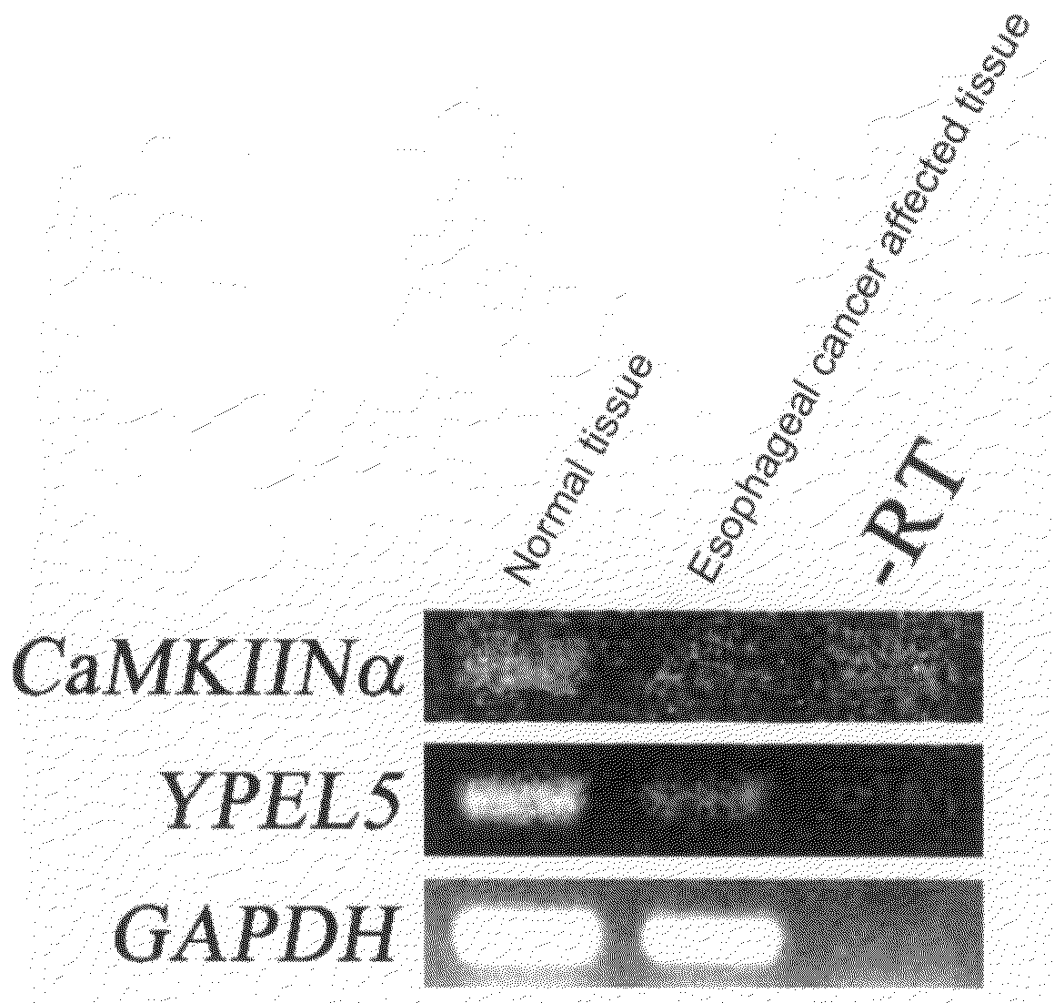
[0661]The tissue of the esophageal cancer lesion in the esophageal tissue and the non-cancerous tissue (normal tissue) of the same esophageal tissue, which tissues were obtained from patients with esophageal cancer, were used as samples. Total RNA was prepared from the above tissues using a Trizol reagent (Invitrogen) in accordance with the manufacturer's recommended protocol. cDNA was synthesized from total RNA using the SuperScript III® First Strand SuperMix, and the cDNA corresponding to 20 ng total RNA was subjected to amplification using TaKaRa Taq® (Takara Bio, Kyoto, Japan). In order to detect the CaMKIINalpha and YPEL5 genes belonging to the group II above, primers (Takara Bio) each consisting of 20 nucleotides corresponding to the gene sequences (SEQ ID NOS: 145 and 158) were subjected to amplification. The composition of the reaction solution was determined in accordance with the provided protocol. The reaction was carried out by the treatment at 95° ...


PUM


Abstract

This invention relates to a composition, kit, or DNA chip comprising polynucleotides and antibodies as probes for detecting, determining, or predicting the presence or metastasis of esophageal cancer, and to a method for detecting, determining, or predicting the presence or metastasis of esophageal cancer using the same.

Description

FIELD OF THE INVENTION[0001]The present invention relates to a composition for diagnosing (i.e., detecting, determining, or predicting) esophageal cancer and metastasis of esophageal cancer, to a method for detecting, determining, or predicting the presence or metastasis of esophageal cancer using the composition, and to a kit for diagnosing the esophageal cancer or the metastasis of the esophageal cancer using the composition.BACKGROUND OF THE INVENTION[0002]The esophagus is a luminal organ that connects the pharynges and the stomach. The major parts thereof are present in the thoracic cavity, and some parts are present in the cervical region and in the abdominal cavity. In the upper portion of the thoracic cavity, the esophagus is located between the trachea and the spine, and it is surrounded by the heart, the aorta, and the lungs in the lower portion. The esophagus delivers food ingested via the mouth to the stomach.[0003]In 2001, the cancer mortality was 238.8 out of 100,000 pa...

Claims


Application Information

Patent Timeline
IPC IPC(8): C40B30/04C12Q1/68G01N33/53C40B40/08
CPCC12Q1/6886G01N33/57407C12Q2600/158C12Q2600/112C12M1/00C12N15/09G01N33/15
Inventor AKIYAMA, HIDEOKOZONO, SATOKOMYOMOTO, AKIRANOMURA, OSAMUNOBUMASA, HITOSHITANAKA, YOSHINORITOMODA, SHIORISHIMADA, YUTAKATSUJIMOTO, GOZOH
Owner TORAY IND INC
Learn More