
Transvalvular intraannular band for valve repair

a transvalvular and intraannular band technology, applied in the field of transannular bands, can solve the problems of loss of ventricular function and geometry, insufficient durability of the valve, and inability to properly function one or more of the valves, and achieve the effect of improving cardiac function

Active Publication Date: 2009-10-22
HEART REPAIR TECH INC
View PDF58 Cites 79 Cited by
  • Summary
  • Abstract
  • Description
  • Claims
  • Application Information

AI Technical Summary

Benefits of technology

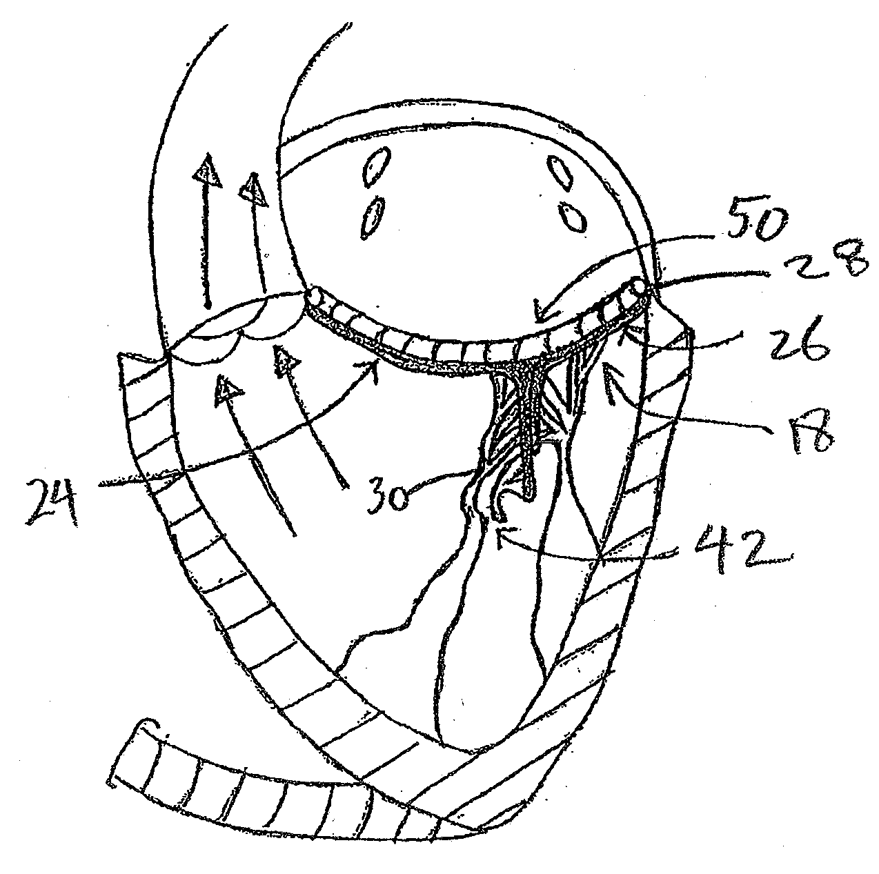
[0017]There is provided in accordance with aspect of the present invention, a transannular band for improving cardiac function. The band comprises an elongate and arcuate body, having a first end, a first anchoring portion located near the first end, and a second end, having a second anchoring portion located near the second end. A central portion is provided, for spanning the flow path of a valve such as a mitral valve. The central portion is displaced transversally from a plane which includes the first end and second end. As implanted, the transverse displacement advances the coaption point of the closed valve in the direction of the ventricle. The first end and second end are configured to be attached to opposing sides of a mitral valve annulus, and the central portion is configured to support the mitral valve leaflets.

Problems solved by technology

Various disease processes can impair the proper functioning of one or more of these valves.
In addition, damage to the ventricle from prior heart attacks (i.e., myocardial infarction secondary to coronary artery disease) or other heart diseases (e.g., cardiomyopathy) can distort the valve's geometry causing it to dysfunction.
The predominant cause of dysfunction of the mitral valve is regurgitation which produces an ineffective cardiac pump function resulting in several deleterious conditions such as ventricular and atrial enlargement, pulmonary hypertension and heart-failure and ultimately death.
Historically, the surgical approach to patients with functional mitral regurgitation was mitral valve replacement, however with certain adverse consequences such as thromboembolic complications, the need for anticoagulation, insufficient durability of the valve, loss of ventricular function and geometry.
These procedures are surgically demanding and require a bloodless and well-exposed operating field for an optimal surgical result.
This method commonly referred to as an edge-to-edge or “Alfieri” repair also has certain drawbacks such as the creation of a double orifice valve and thereby reducing the effective orifice area.
However, it still remains to conduct an annuloplasty procedure, which has not yet been resolved by a catheter technique and therefore is to be performed by conventional surgery, which makes the method impractical.

Method used


Image

  • Transvalvular intraannular band for valve repair
  • Transvalvular intraannular band for valve repair
  • Transvalvular intraannular band for valve repair

Examples


Embodiment Construction

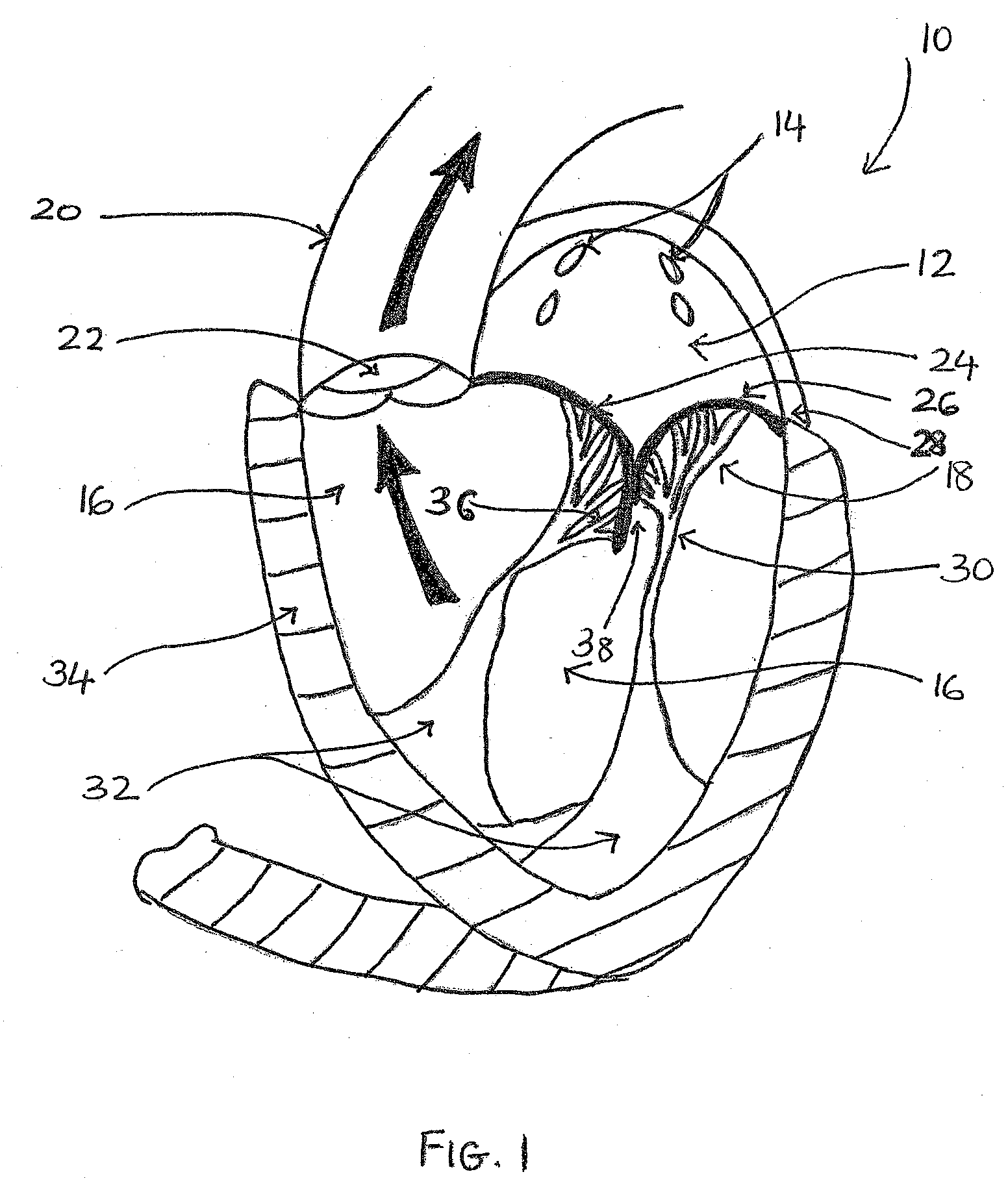
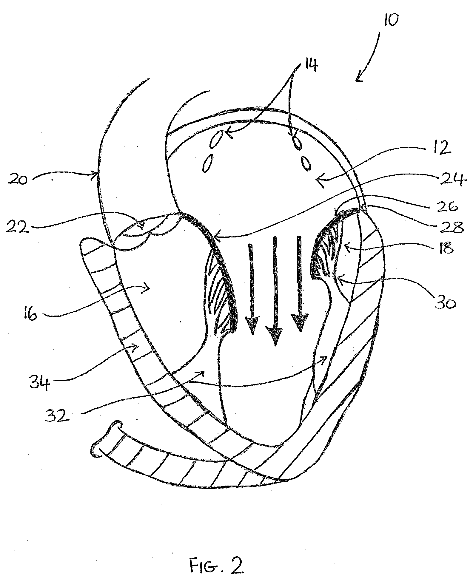
[0060]FIG. 1 illustrates a cross-sectional view of the heart 10 with a normal mitral valve 18 in systole. As illustrated, the heart 10 comprises the left atrium 12 which receives oxygenated blood from the pulmonary veins 14 and the left ventricle 16 which receives blood from the left atrium 12. The mitral valve 18 is located between the left atrium 12 and left ventricle 16 and functions to regulate the flow of blood from the left atrium 12 to the left ventricle 16. During ventricular diastole, the mitral valve 18 is open which allows blood to fill the left ventricle 16. During ventricular systole, the left ventricle 16 contracts, which results in an increase in pressure inside the left ventricle 16. The mitral valve 18 closes when the pressure inside the left ventricle 16 increases above the pressure within the left atrium 12. The pressure within the left ventricle 16 continues increasing until the pressure within the left ventricle 16 exceeds the pressure within the aorta 20, which...


PUM


Abstract

Mitral valve prolapse and mitral regurgitation can be treating by implanting in the mitral annulus a transvalvular intraannular band having an elongate and arcuate body. The elongate and arcuate body has a first end, a first anchoring portion located proximate the first end, a second end, a second anchoring portion located proximate the second end, and a central portion. The central portion is displaced from the plane containing the first end and the second end. The transvalvular band is positioned so that it extends transversely across a coaptive edge formed by the closure of the mitral valve leaflets and the central portion is displaced towards the left ventricle relative to the first anchoring portion and the second anchoring portion. The ventricular direction displacement moves coaption to an earlier point in the cardiac cycle.

Description

BACKGROUND OF THE INVENTION[0001]1. Field of the Invention[0002]Embodiments of the present invention relate generally to treatment of mitral or tricuspid valve prolapse and mitral regurgitation, and more specifically, relate to the use of a transannular band to treat mitral valve prolapse and mitral regurgitation.[0003]2. Description of the Related Art[0004]The heart is a double (left and right side), self-adjusting muscular pump, the parts of which work in unison to propel blood to all parts of the body. The right side of the heart receives poorly oxygenated (“venous”) blood from the body from the superior vena cava and inferior vena cava and pumps it through the pulmonary artery to the lungs for oxygenation. The left side receives well-oxygenated (“arterial”) blood from the lungs through the pulmonary veins and pumps it into the aorta for distribution to the body.[0005]The heart has four chambers, two on each side—the right and left atria, and the right and left ventricles. The at...

Claims


Application Information

Patent Timeline
Patent Type & Authority Applications(United States)
IPC IPC(8): A61F2/24
CPCA61F2/2454
Inventor SUBRAMANIAN, VALAVANUR A.
Owner HEART REPAIR TECH INC
Learn More