
Bioactive peptide for cell adhesion

a technology of cell adhesion and bioactive peptide, which is applied in the field of bioactive peptide for cell adhesion, can solve the problems of inability to commercially exploit the intact molecule as a cellular adhesive, and the size of the intact molecule does not allow for efficient and cost-effective commercial production

Inactive Publication Date: 2009-09-24
UNIVERSITY OF DELAWARE
View PDF4 Cites 1 Cited by
  • Summary
  • Abstract
  • Description
  • Claims
  • Application Information

AI Technical Summary

Benefits of technology

The patent describes an implantable device that can stick to eukaryotic cells, which are the type of cells that make up most of the body. The device is made by coating a scaffold with a specific protein that the cells can stick to. The device can be used to repair tissue or organs in the body. The process involves putting the scaffold in the body and adding cells to it, allowing the cells to stick to the scaffold and grow on it. This can help to create new tissue or organs that can replace damaged ones.

Problems solved by technology

Although it is known that perlecan is involved in cell adhesion, the intact molecule is too large to exploit commercially as a cellular adhesive.
Its size does not allow for efficient and cost effective commercial production.

Method used


Image

  • Bioactive peptide for cell adhesion
  • Bioactive peptide for cell adhesion
  • Bioactive peptide for cell adhesion

Examples


example 1

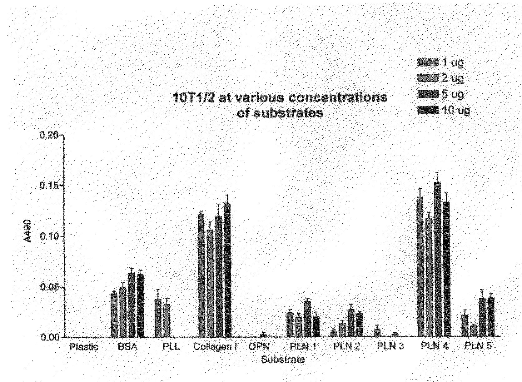
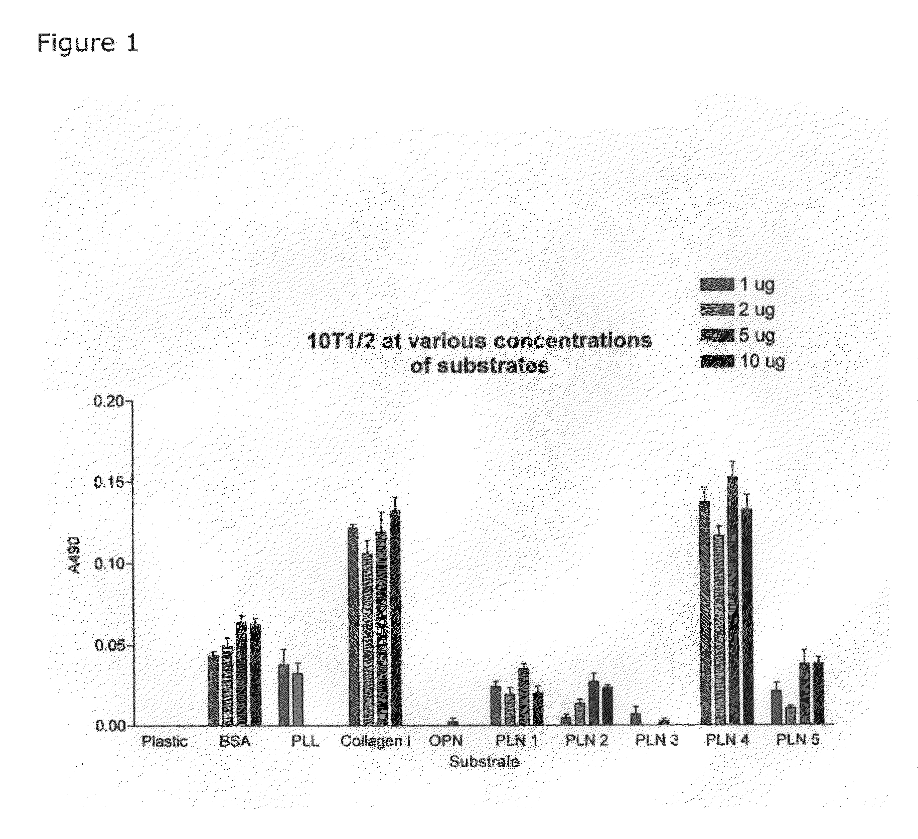
[0060]FIG. 1 shows the results of studies comparing adhesion of multipotent mouse mesenchymal stem cells, 10T1 / 2, to uncoated plastic and to plastic coated with various substrates. Adhesion is measured by the cell proliferation assay CELLTITER 96® AQueous Non-Radioactive Cell Proliferation Assay (MTS) sold by Promega, Inc. The CELLTITER 96® AQueous Non-Radioactive Cell Proliferation Assay is a calorimetric method for determining the number of viable cells in proliferation, cytotoxicity or chemosensitivity assays. The CELLTITER 96® AQueous Non-Radioactive Cell Proliferation Assay is composed of solutions of a tetrazolium compound (3-(4,5-dimethylthiazol-2-yl)-5-(3-carboxymethoxyphenyl)-2-(4-su-lfophenyl)-2H-tetrazolium, inner salt; MTS) and an electron coupling reagent (phenazine methosulfate) PMS. MTS is bioreduced by cells into a formazan that is soluble in tissue culture medium. The absorbance of the formazan at 490 nm can be measured directly from 96 well assay plates without add...

example 2

[0063]Cell Adhesion Assay

[0064]Concentrated stocks of rat-tail type I collagen were diluted to designated concentrations in 0.02 N acetic acid and coated overnight on MAXISORP® 96 well plates at room temperature (RT). Poly-L-lysine (PLL), heat-denatured bovine serum albumin (BSA), the peptide of SEQ ID NO:1 conjugated to BSA (indicated in FIGS. 4-7 as “PLN4”) and a scrambled peptide from domain IV of perlecan having the amino acid sequence TGKSVGGSLIWPVRGQH (SEQ ID NO:4) conjugated to BSA (indicated in FIGS. 4-7 as “PLN4*”) were diluted in 1×phosphate buffered saline (PBS) and also coated overnight on MAXISORP® 96 well plates at RT. The following day, the plates were washed three times with 1×PBS to remove any residue and blocked with 1% heat-denatured BSA for 1 hr at 37° C. Following blocking, plates were washed once with 1×PBS. Cells from various cell lines were harvested with 0.5 mM EDTA and collected by centrifugation. Pelleted cells were resuspended in serum-free RPMI 1640 and ...


PUM

PropertyMeasurementUnit
molecular weightaaaaaaaaaa

Abstract

Implantable devices for adhering eukaryotic cells and devices providing a substrate for eukaryotic cell growth and / or differentiation in vitro are described. Each device comprises a scaffold that is coated with a protein comprising an adhesive polypeptide that has at least 90% sequence identity to a specific peptide sequence within domain IV of perlecan. The devices adhere epithelial cells, epithelial stem cells, mesenchymal stem cells, and osteoblasts.

Description

CROSS REFERENCE TO RELATED APPLICATIONS[0001]This is a divisional application of U.S. application Ser. No. 10 / 363,376 (status: pending), filed Jul. 7, 2003, which is a National Phase Application of PCT Application Ser. No. US01 / 026640, filed Aug. 27, 2001, which claims the benefit of U.S. Provisional Application Ser. No. 60 / 229,222, filed Aug. 31, 2000, and U.S. Provisional Application Ser. No. 60 / 309,209, filed Aug. 1, 2001, all of which are incorporated herein, in entirety, by reference.GOVERNMENT INTERESTS[0002]This invention was made with government support under Grant Nos. HD25235; DE13542, RR16458, and CA098912, Project 2, awarded by the National Institutes of Health. The government has certain rights in this invention.BACKGROUND OF THE INVENTION[0003]Perlecan is a heparan sulfate proteoglycan (Noonan, D. M. (1991) J. Biol. Chem. (266) 34: 22939-22947). The proteoglycan family contains more than 30 members that perform an assortment of biological functions and are widely distr...

Claims


Application Information

Patent Timeline
Patent Type & Authority Applications(United States)
IPC IPC(8): C12N5/06A61K38/00A61L27/34A61L27/38C07H21/04C07K14/47
CPCA61K38/00A61L27/34A61L27/3813A61L27/3843C07K14/4725C07H21/04C08L89/00
Inventor FARACH-CARSON, MARY C.CARSON, DANIEL D.SAFRAN, JEFFREY B.
Owner UNIVERSITY OF DELAWARE
Learn More