Method for treating brain ischemic injury through transplantation of human umbilical mesenchymal stem cells
a technology of stem cells and brain ischemic injury, applied in the field of stem cells, can solve the problems of stroke damage, irreversible long-term sensorimotor deficit, neuronal injury, etc., and achieve the effect of restoring stroke damage completely and avoiding stroke damag
- Summary
- Abstract
- Description
- Claims
- Application Information
AI Technical Summary
Benefits of technology
Problems solved by technology
Method used
Image
Examples
example 1
The Infarct Volume of Stroke Rats After MCAO Surgery
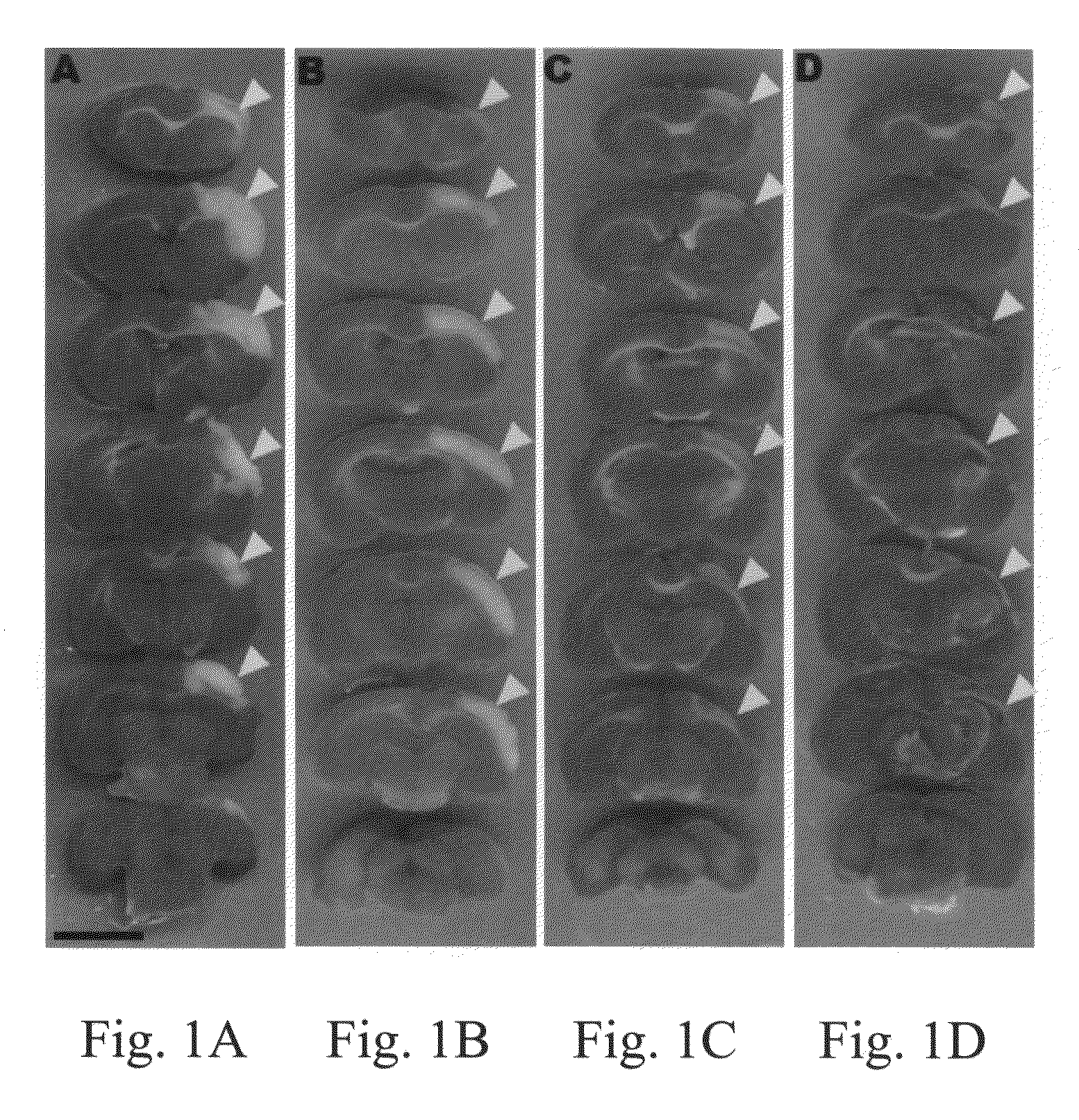
[0056]Using the histological examination and immunochemical analysis of grafted brain cryosections explained above, the ranges of damaged cortex in stroke rats were examined. Individual groups of stroke rats were sacrificed at day 1, 8, 15, or 29 after MCAO surgery. These rats were used for TTC stain, a common method in stroke rodent study (Bederson, J. B., et al., Stroke 1986, 17: 1304-1308). Normal brain tissue was stained for red color, and damaged areas appeared as white color. FIG. 1A showed that quantitative analysis of infarcted brain volume demonstrated that the damaged cortex was inflamed and edemic, resulting in a significant increase of volume at 1 day after MCAO (p3). At day 8, as shown in FIG. 1B, after MCAO, the edema of the inflamed cortex was alleviated, and the volume of damaged brain was 154.29±6.52 mm3. FIG. 1C showed that the infarcted cortex started to express atrophy compared with the contralateral normal cort...
example 2
HUMSCs Transplantation Reduces Damage Area of Stroke Rat Brain
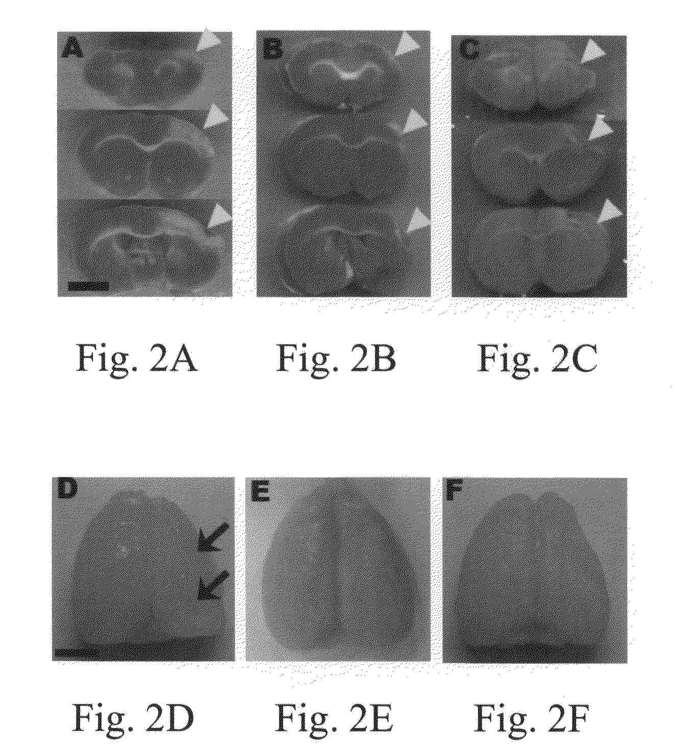
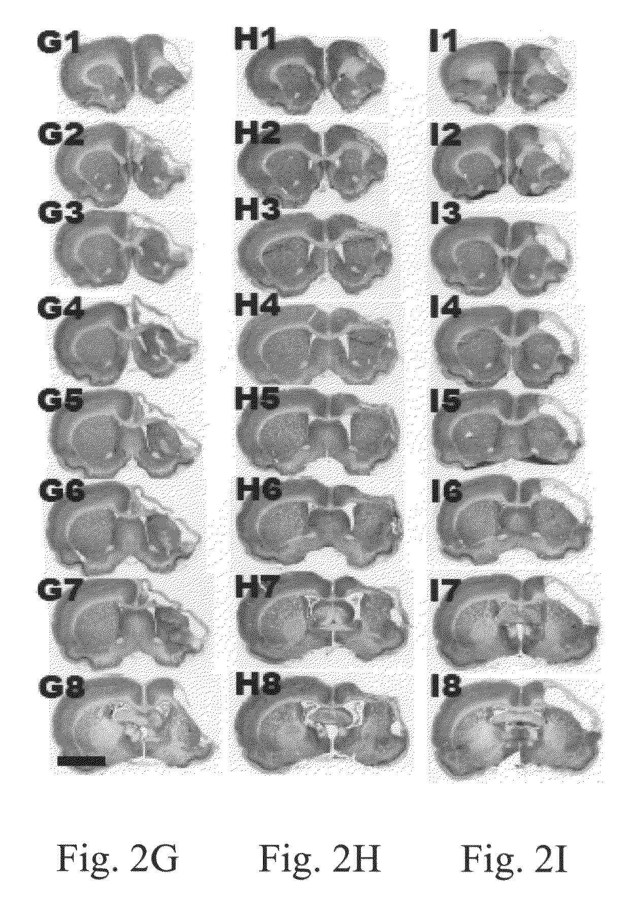
[0057]At day 8 after transplantation, the changes of ischemic cortices using TTC stain were examined for three different groups (Control, Stem cell, and NCM 6D). FIGS. 2A-2C illustrated that the damaged areas of stroke rat brains in the stem cell group and the NCM 6D group are significantly reduced as compared with the control group. At day 36 after transplantation, representative brain cortical expression from stroke rats in each of the control, stem cell and NCM 6D groups is shown in FIGS. 2D-2F, respectively. The appearance of atrophy in the control group was more serious in the control group (FIG. 2D) than in the stem cell (FIG. 2E) and the NCM 6D (FIG. 2F) groups. FIGS. 2G-2I showed the similar patterns from rostral to caudal slices of brain stained with cresyl-violet stain. The brain slices shown in FIG. 2G1-2G8 from the control group displayed harsh damage in the infarct cortex and even in the basal ganglion.
[0058]...
example 3
HUMSCs Transplantation Recovers Neurological Behavior Deficits
[0059]The effects of HUMSCs transplantation on functional recovery were examined for the cylinder and rotarod tests. These tests were performed before and after MCAO, and at days 1, 4, 8, 15, 22, 29, and 36 after transplantation. In the cylinder test as shown in FIG. 4A, normal rats before MCAO used both forelimbs at almost same percentage, displayed as 50%. After MCAO (day −1), contralateral forelimb usage percentage of the control group treated without stem cells decreased to 10.38±2.65%, and increased to 21.44±3.55% after treatment with PBS. But compared to the normal value, the tendency still showed a statistically significant difference until 36 days (p36 days, just as the control group did. As shown by the data, one day after MCAO, usage of contralateral forelimb decreased to 19.84%, about 30.13% of the normal value. However, usage of the contralateral forelimb recovered to 65.95%, about 67.88% of the normal value a...
PUM
 Abstract
Description
Claims
Application Information
 - R&D
- Intellectual Property
- Life Sciences
- Materials
- Tech Scout
- Unparalleled Data Quality
- Higher Quality Content
- 60% Fewer Hallucinations
Browse by: Latest US Patents, China's latest patents, Technical Efficacy Thesaurus, Application Domain, Technology Topic, Popular Technical Reports.
© 2025 PatSnap. All rights reserved.Legal|Privacy policy|Modern Slavery Act Transparency Statement|Sitemap|About US| Contact US: help@patsnap.com


