
Biosensor

a biosensor and sensor technology, applied in the field of biosensors, can solve the problems of inability to detect, difficult to achieve, and difficult to obtain analysis results,

Inactive Publication Date: 2009-04-02
TDK CORPARATION
View PDF2 Cites 5 Cited by
  • Summary
  • Abstract
  • Description
  • Claims
  • Application Information

AI Technical Summary

Benefits of technology

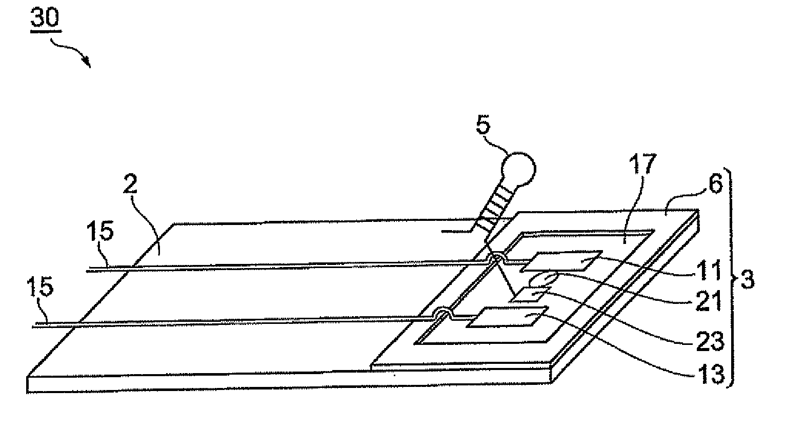
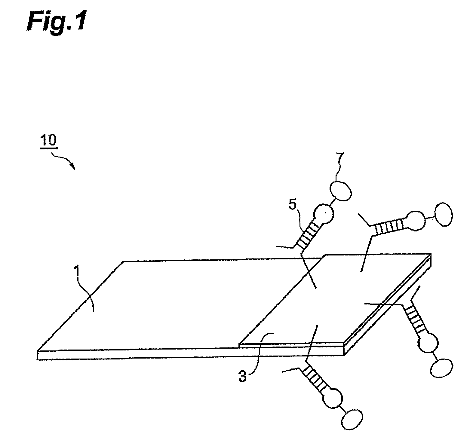
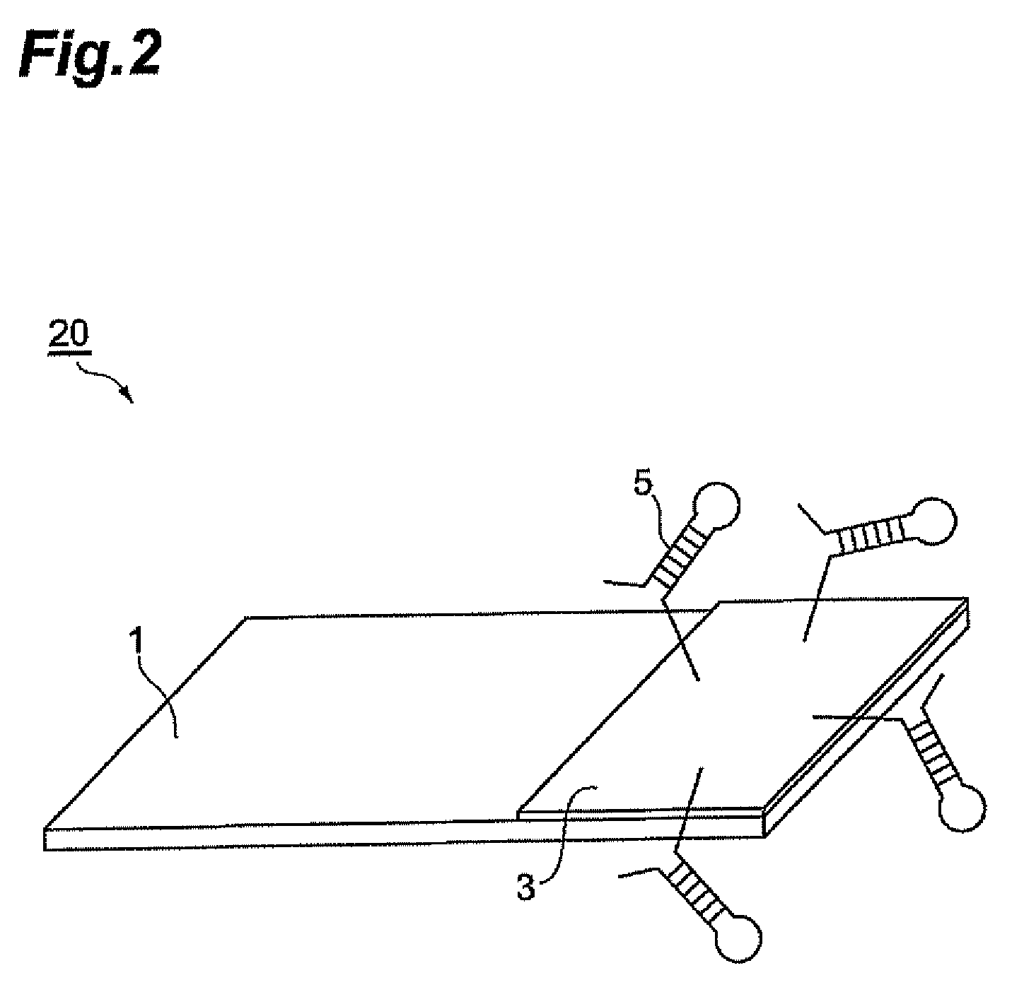
[0030]Since the substrate recognition element in the biosensor of the invention is resistant to enzymolysis and drying, the biosensor is characterized in that its detection sensitivity for target bacterium is not impaired depending on the storage condition of the biosensor or depending on the measuring sample. In addition, the substrate recognition element of the biosensor of the invention does not contain agents that are harmful to the human body, and therefore the substrate recognition element can be directly inserted into the mouth to allow direct measurement of intraoral bacterium in saliva.
[0031]Moreover, since the substrate recognition element of the biosensor of the invention contains an aptamer(s) that binds with a protein comprising any of the amino acid sequences as set forth in SEQ ID NO: 1-25, it is possible to accomplish specific detection of Streptococcus mutans, Streptococcus sobrinus or Lactobacillus acidophilus.

Problems solved by technology

Detection of bacterium that is invisible to the naked eye, however, currently involves specimen extraction, identification by culturing in selective medium, and detection of the bacterium by antibody-based ELISA or Western blotting methods, and effort and time are required to obtain the analysis results.
Moreover, while biosensors comprising monoclonal antibody immobilized on the substrate recognition element can in principle detect analytes in a shorter time compared to ELISA or Western blotting methods, the antibody immobilized on the substrate recognition element is easily affected by drying and by proteases during storage and use of the biosensor, and therefore the detection sensitivity is often significantly reduced.
Such biosensors are therefore associated with problems of stability.
Also, biosensors having monoclonal antibody immobilized on the substrate recognition element often employ substances that are harmful to the human body to maintain antibody stability, therefore the substrate recognition elements of such biosensors cannot be directly inserted into the mouth for detection of intraoral bacterium, and it has been necessary to perform indirect measurement of samples such as saliva in separate containers.

Method used


Image

  • Biosensor
  • Biosensor
  • Biosensor

Examples


example 1

[0068]Screening of Aptamers that Bind Specifically to Streptococcus mutans

[0069]Streptococcus mutans pac (SEQ ID NO: 13) was selected as a bacterial cell surface molecule specifically found on Streptococcus mutans, and aptamers that specifically bind to this protein were screened.

[0070]First, the nucleotide sequence of Streptococcus mutans pac represented by SEQ ID NO: 13 of the Sequence Listing, as the aptamer-binding site, was processed with a computer evolution program to generate 10 generations of nucleotide sequences on the computer, and the nucleotide sequences were used as candidate Streptococcus mutans-binding aptamers. The computer evolution program was written with Visual Basic using a common genetic algorithm with reference to the one by Ikebukuro Nucleic Acids Res., 2005, Vol. 33, e108).

[0071]Next, oligo DNA consisting of the candidate nucleotide sequences was chemically synthesized, and those exhibiting specific binding for Streptococcus mutans were recovered as aptame...

example 2

[0073]Detection of Streptococcus mutans with Aptamer(s)

[0074]Detection of Streptococcus mutans by ELISA was attempted, using S. mutans-binding aptamer(s) instead of anti-Streptococcus mutans antibody.

[0075]First, Streptococcus mutans was suspended in PBS and cell suspensions of different concentrations (1×10⁴, 1×10⁵, 1×10⁶, 1×10⁷, 1×10⁸ and 1×10⁹ CFU / mL) were prepared. The prepared cell suspensions were added in 100 μL aliquots to each well of a poly-L-lysine-coated 96-well plate, and were incubated for a prescribed period of time to fix the cells at the bottoms of the wells.

[0076]To each of the cell-fixed wells there was added 50 μg of the biotin-labeled S. mutans-binding aptamer(s) prepared in Example 1, and after incubation at 25° C. for 1 hour, they were rinsed with PBS to remove the biotin-labeled S. mutans-binding aptamer(s) that had not bound to the cells.

[0077]Next, HRP-labeled steptavidin was added to each well, and after incubation at 25° C. for 1 hour, each well was thoro...

example 3

[0087]Detection of Streptococcus mutans with Biosensor Comprising S. mutans-Binding Aptamer(s) in Substrate Recognition Element (Electrochemical Method)

[0088]A sensor was constructed with electrolyte solution in a 3-electrode type electrochemical cell comprising a working electrode having the S. mutans-binding aptamer(s), alkanethiol and ferrocene immobilized on the electrode, and a counter electrode and a reference electrode, and was used to detect Streptococcus mutans in a test sample.

[0089]First, biotin-labeled alkanethiol was bonded to the surface of the gold electrode and the gold electrode was rinsed with PBS, and then the avidin-labeled ferrocene was bound to the alkanethiol by biotin-avidin reaction, the gold electrode was again rinsed with PBS, and finally the biotin-labeled S. mutans-binding aptamer(s) prepared in Example 1 was bound to the ferrocene by biotin-avidin reaction to form the working electrode (substrate recognition element).

[0090]A 3-electrode type electrochem...


PUM

PropertyMeasurementUnit
fluorescent wavelengthaaaaaaaaaa

Abstract

The invention provides a biosensor comprising a microbe-binding aptamer(s) in the substrate recognition element. It is possible to obtain a stabilized biosensor wherein the detection sensitivity for target microbe (target bacterium) is not impaired depending on the storage condition or measuring sample, and target bacterium in a body fluid can be directly measured by insertion of the substrate recognition element of the biosensor.

Description

RELATED APPLICATION[0001]This application claims priority to Japanese patent application 2007-252148 filed on Sep. 27, 2007, which is herein incorporated by reference in its entirety.BACKGROUND OF THE INVENTION[0002]1. Field of the Invention[0003]The present invention relates to a biosensor.[0004]2. Related Background Art[0005]“Biosensor” is a general term for chemical sensors that utilize biological molecular recognition mechanisms, and known types of biosensors include enzyme sensors, immunosensors, microbiological sensors, ion channel sensors and the like.[0006]Biosensors accomplish detection of analytes by converting a change (for example, a substance change, color change, heat absorption or release, mass change or the like) produced-by interaction between an enzyme, antibody, microbe, ion channel or the like that is immobilized on a substrate recognition element, and its substrate (the analyte), into a signal that is detectable using a signal converter (an electrode, light rece...

Claims


Application Information

Patent Timeline
Patent Type & Authority Applications(United States)
IPC IPC(8): G01N33/569
CPCG01N33/5438G01N2333/335G01N33/56955G01N33/56944
Inventor ONO, SHIZUKOTAKAHASHI, YAYOI
Owner TDK CORPARATION
Learn More