Miniaturized lateral flow device for rapid and sensitive detection of proteins or nucleic acids

Inactive Publication Date: 2009-02-19
LOS ALAMOS NATIONAL SECURITY
View PDF48 Cites 62 Cited by
  • Summary
  • Abstract
  • Description
  • Claims
  • Application Information

AI Technical Summary

Benefits of technology

[0009]The LFM technology and LFM devices of the invention offer many of the advantages of microarray technology yet retain the simplicity of lateral flow-based platforms. The miniaturization of lateral flow nucleic acid detection achieved by the present invention offers reduced reagent use, femtomole sensitivity, excellent linear dynamic range, and rapid detection. Moreover, the small feature sizes of capture oligonucleotides renders the potential information capacity of the platform comparable to more traditional spotted fluorescence microarrays as well as improving sensitivity. The LFM devices exemplified herein enable analytes to be detected within 10 seconds from the time of sample introduction to the LFM device. Sample volumes may be as low as about 10 microliters, significantly reducing assay costs and ameliorating reagent storage logistics. Additionally, the miniaturization of lateral flow opens the door to highly multiplexed assays, allowing many proteins or nucleic acids to be detected in a single assay.
[0010]Coupled with an isothermal amplification technique, LFM provides a facile means of rapidly detecting nucleic acid targets while circumventing hardware requirements for fluorescence detection and PCR thermocycling.
[0011]The power of LFM is demonstrated in the Examples, infra. More specifically, Example 8 illustrates the utility of the lateral flow microarray (LFM) approach for sensitive detection and discrimination of closely related microbial signatures when present as minority sequences in complex nucleic acid mixtures, using an assay based on the nonsense mutation in the plcR gene of B. anthracis, that is absent in the near phylogenetic neighbors B. thuringiensis and B. cereus (27,28). The results demonstrate that LFMs, making use of stable detection reagents suitable for dry storage, can be used to detect as little as 250 amol analyte within 2 minutes of sample addition. The miniaturization of lateral flow detection decreases reagent consumption and sample-to-answer times while increasing the potential information capacity of the platform to enable the development of highly multiplexed nucleic acid detection assays.
[0013]In some embodiments, the first sequence and second sequence of the target nucleic acid are adjacent within 2 bases, in order to take advantage of “base stacking” hybridization stability.
[0015]In some embodiments, the first sequence and second sequence of the target nucleic acid are adjacent within 2 bases, in order to take advantage of “base stacking” hybridization stability.

Problems solved by technology

While nucleic acid-based assays for pathogen detection and identification offer sensitivity, specificity and resolution, they are relatively elaborate and often costly, limiting their utility for point-of-care diagnostics and deployment under field conditions where a supporting laboratory infrastructure is limited or absent.
Retaining assay sensitivity, while circumventing requirements for thermocyclers and fluorescence detection hardware, remains a significant challenge.
Nonetheless, the reliance of this technology on costly instrumentation for high-resolution fluorescence signal transduction severely limits the utility of microarrays for field applications where a laboratory infrastructure is limited or unavailable.
Though microarray hybridization times as short as 500 seconds have been reported (9), such methods employ relatively elaborate microfluidic designs that remain reliant upon fluorescent detection and do not address the need for low cost, easily manufactured devices that can be used without costly supporting instrumentation.
Unfortunately, the utility of lateral flow detection in the context of a PCR-based assay is severely limited by the fact that reliance on thermocycling hardware largely negates the potential benefit of the otherwise highly simplified lateral flow platform.

Method used


Image

  • Miniaturized lateral flow device for rapid and sensitive detection of proteins or nucleic acids
  • Miniaturized lateral flow device for rapid and sensitive detection of proteins or nucleic acids
  • Miniaturized lateral flow device for rapid and sensitive detection of proteins or nucleic acids

Examples


example 1

Detection of DNA Hybridization Over Broad Range of Capture Oligonucleotide Deposition Concentrations on DNA Dipstick

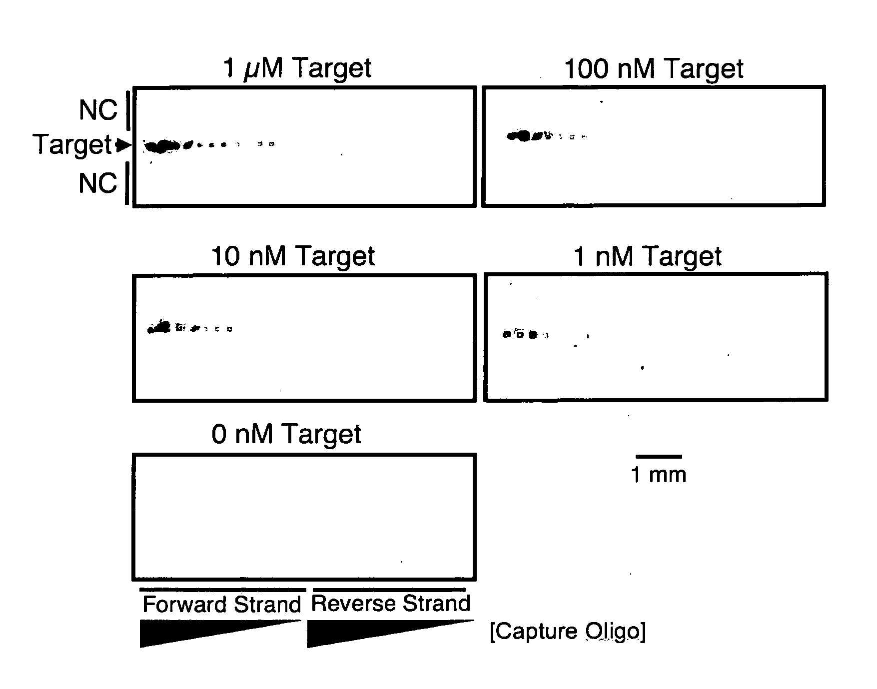
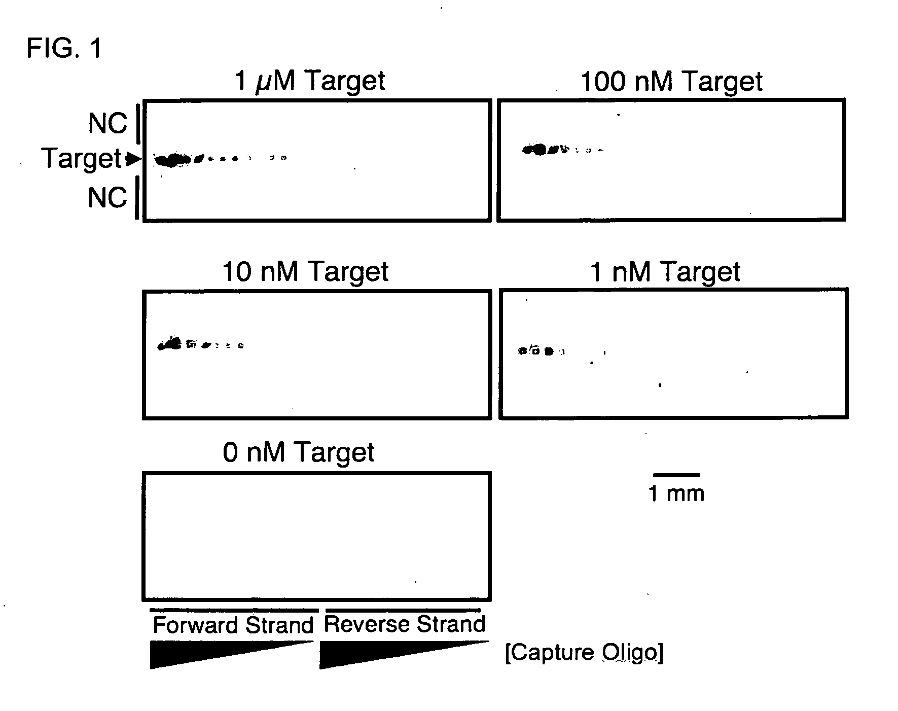
[0097]DNA dipstick microarrays were fabricated at a density of 36 features per mm2 using varying concentrations of capture oligonucleotide, as indicated In FIG. 1. Printing solutions of capture oligonucleotide at 200 μM, 100 μM, 50 μM, 25 μM, 12.5 μM, 6.25 μM, and 3.125 μM were prepared and patterned on to lateral flow membranes. The resulting DNA dipstick microarrays were introduced to 100 μl of synthetic target DNA at the indicated concentration of 1 μM, 100 nM, 10 nM, 1 nM, and 0 nM corresponding to 100 μmol, 10 μmol, 1 μmol, 100 fmol, and 0 fmol target molecules respectively. Capture of 100 fmol target molecule was apparent at capture oligonucleotide printing concentrations as low as 12.5 μM (FIG. 1). However, the most sensitive detection was obtained at higher capture oligonucleotide printing concentrations of 200 μM. Subsequent DNA dipsticks and DNA dipstick micr...

example 2

Detection of Single and Multiple SS-DNA Species

[0098]The following example demonstrates sensitive detection of single-stranded DNAs using hybridization-based capture and dyed-microsphere calorimetric detection.

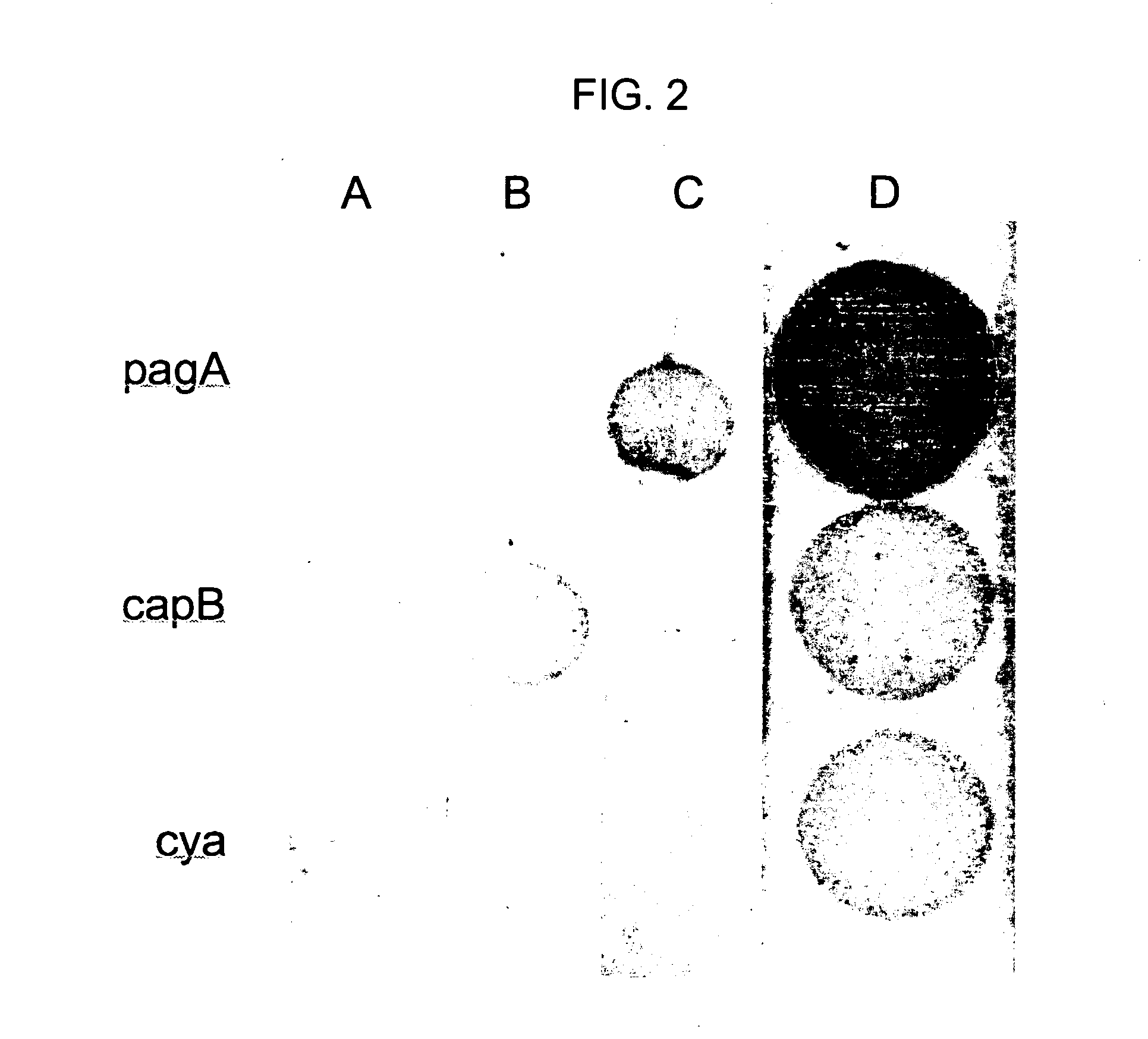
[0099]Sequences derived from the B. anthracis pagA, capB and cya genes were used to demonstrate multiplexed detection. DNA Dipsticks were patterned with capture sequences for the detection of fragments of three key virulence factors of B. anthracis.

[0100]The capture sequences used were:

pagD:[SEQ ID NO: 7]5′- GCAGGATTTAGTAATTCGAATTTTTTTTTTTTTTT-3′;cyaD908:[SEQ ID NO: 8];5′ TGGTACTAAACCTGAAGCTTTTTTTTTTTTTTTTT 3′;andcapD:[SEQ ID NO: 9]5′-TACATGGTCTTCCCAGATAATTTTTTTTTTTTTTT-3′.

0.35 μm dyed COOH microspheres (Spherotech, Inc.) were coupled using EDAC and standard protocols to:

[SEQ ID NO: 10]5′ amine-C12-TTTTTTTTTTTTTTTTTTCAGAAGAATTCTTACGAAAATTTGAT 3′ for capB detection,[SEQ ID NO: 11]5′ amine-C12-TTTTTTTTTTTTTTTTTCTTTGATATTGGTGGGAGTGTATC for pagA detection;and[SEQ ID NO: 12]5′ ami...

example 3

Sensitivity and Detection Time for DNA Dipstick and DNA Dipstick Microarray

[0102]The sensitivity and time required to detect nucleic acids on DNA dipsticks and DNA dipstick microarrays were evaluated using synthetic target molecules for which exact concentrations could be determined.

[0103]Dipsticks were fabricated by manual deposition of capture oligonucleotides onto membrane strips of approximately 160 to 275 mm2 surface area, features sizes were ˜2-3 mm in diameter. Dipstick microarrays were printed using microarray fabrication robotics to pattern membrane strips of approximately 60 mm2 surface area such that feature sizes were 300-600 μm in diameter. DNA dipsticks were challenged with 400 μl of synthetic target molecule in the presence of appropriate detection microspheres. Typical lateral flow time for these strips was approximately 45 minutes from sample introduction to complete transport of the sample through the dipstick matrix. Dilution series experiments revealed the sensit...


PUM

PropertyMeasurementUnit
Lengthaaaaaaaaaa
Lengthaaaaaaaaaa
Pore sizeaaaaaaaaaa

Abstract

The invention provides miniaturized lateral flow chromatographic and lateral flow chromatographic microarray devices (LFM). The miniaturization of lateral flow nucleic acid detection achieved by the present invention offers reduced reagent use, femtomole sensitivity, excellent linear dynamic range, and rapid detection. Moreover, the small feature sizes of capture oligonucleotides renders the potential information capacity of the platform comparable to more traditional spotted fluorescence microarrays as well as improving sensitivity. The LFM devices exemplified herein enable analytes to be detected within 10 seconds from the time of sample introduction to the LFM device. Sample volumes may be as low as about 10 microliters, significantly reducing assay costs and ameliorating reagent storage logistics. Additionally, the miniaturization of lateral flow opens the door to highly multiplexed assays, allowing many proteins or nucleic acids to be detected in a single assay.

Description

RELATED APPLICATIONS[0001]This patent application claims the benefit of the filing date of U.S. Provisional patent application No. 60 / 839,537 filed Aug. 22, 2006 and Provisional patent application No. 60 / 925,210 filed Apr. 18, 2007 under 35 U.S.C. 119(e).STATEMENT AS TO RIGHTS TO INVENTIONS MADE UNDER FEDERALLY SPONSORED RESEARCH OR DEVELOPMENT[0002]This invention was made with government support under Contract No. DE-AC52-06NA25396, awarded by the United States Department of Energy. The government has certain rights in this invention.BACKGROUND OF THE INVENTION[0003]The threat presented by biological weapons, global heath care issues and emerging diseases of natural origin lend urgency to the development of rapid, field-deployable pathogen detection and diagnostic tools (1,2). Ideally, to be of general field utility, a diagnostic device must be capable of sensitive and specific pathogen detection while retaining simplicity of use and independence from complex laboratory instrumenta...

Claims


Application Information

Patent Timeline
IPC IPC(8): C12Q1/68G01N30/00C40B60/12
CPCC12Q1/6834C12Q2565/625C12Q1/6837G01N33/523
Inventor CARY, ROBERT B.
Owner LOS ALAMOS NATIONAL SECURITY
Why Eureka
Try Eureka