Ultrasonic imaging apparatus and image processing apparatus

a technology of ultrasonic imaging and image processing, applied in the field of ultrasonic imaging apparatus and image processing apparatus, can solve the problems of not being able to achieve a final stereoscopic display, unable and not being able to achieve stereoscopic display in a limited region. , to achieve the effect of less nois

Active Publication Date: 2009-01-29
GE MEDICAL SYST GLOBAL TECH CO LLC
View PDF29 Cites 26 Cited by
  • Summary
  • Abstract
  • Description
  • Claims
  • Application Information

AI Technical Summary

Benefits of technology

[0010]An ultrasonic imaging apparatus according to a first aspect of the invention is characterized by including a 3D tomographic image data capturing device which captures 3D tomographic image data from a 3D region inside a subject, a region-of-interest setting device which sets a 3D region of interest corresponding to the 3D region in an image memory that stores the 3D tomographic image data, a surface image extracting device which extracts data on a surface image, in the 3D region of interest, of a massive tissue included in the 3D region using the 3D tomographic image data, a stereoscopic display generating device which generates stereoscopic display data to display the surface image data stereoscopically, and a display device which displays the stereoscopic display data, wherein the region-of-interest setting device displays a 2D tomographic image of a 2D region including the massive tissue on the display device, allows manual setting of a marker indicating a periphery of the massive tissue in the 2D tomographic image, and generates the 3D region of interest with the marker in the vicinity of the center according to data on the marker's position in the 2D tomographic image.
[0017]According to the fourth aspect of the invention, an operator easily grasps the main content of the 3D tomographic image data from the displayed tomographic image data on the three orthogonal sectional planes.
[0025]According to the eighth aspect of the invention, 3D tomographic image data is captured more quickly by the 2D probe array.
[0027]According to the ninth aspect of the invention, a massive tissue boundary included in the 2D tomographic image is easily specified.
[0037]According to the fourteenth aspect of the invention, a surface image and a stereoscopic display of the surface image are clear with less noise.
[0041]In one embodiment, a 2D region of interest is set so that the massive tissue boundary is included in the 3D region of interest to the maximum extent, and therefore an optimum stereoscopic image is obtained from the beginning and the trouble of setting a 2D region of interest repeatedly is saved and an optimum surface image is easily and quickly displayed.

Problems solved by technology

However, according to the above background art, the position of the stereoscopically displayed surface image of the massive tissue is not well balanced in the 3D region of interest.
Since the purpose of a stereoscopic display of a surface image is to enable one to grasp an overall picture of a massive tissue easily, a stereoscopic display only in a limited region is not desirable.
Therefore, the operator has to set a 2D region of interest repeatedly so that the boundary constituting the surface of the massive tissue spreads all over the 3D region of interest, which means that it takes much time and labor to obtain a final stereoscopic display.
In this case, if it should take time and labor to make a stereoscopic display of the fetus after capturing 3D tomographic image data, it would be frustrating and undesirable for both the operator and the subject.

Method used


Image

  • Ultrasonic imaging apparatus and image processing apparatus
  • Ultrasonic imaging apparatus and image processing apparatus
  • Ultrasonic imaging apparatus and image processing apparatus

Examples


Embodiment Construction

[0054]Next, embodiments of the invention as an ultrasonic imaging apparatus will be described referring to the accompanying drawings. It does not limit the invention.

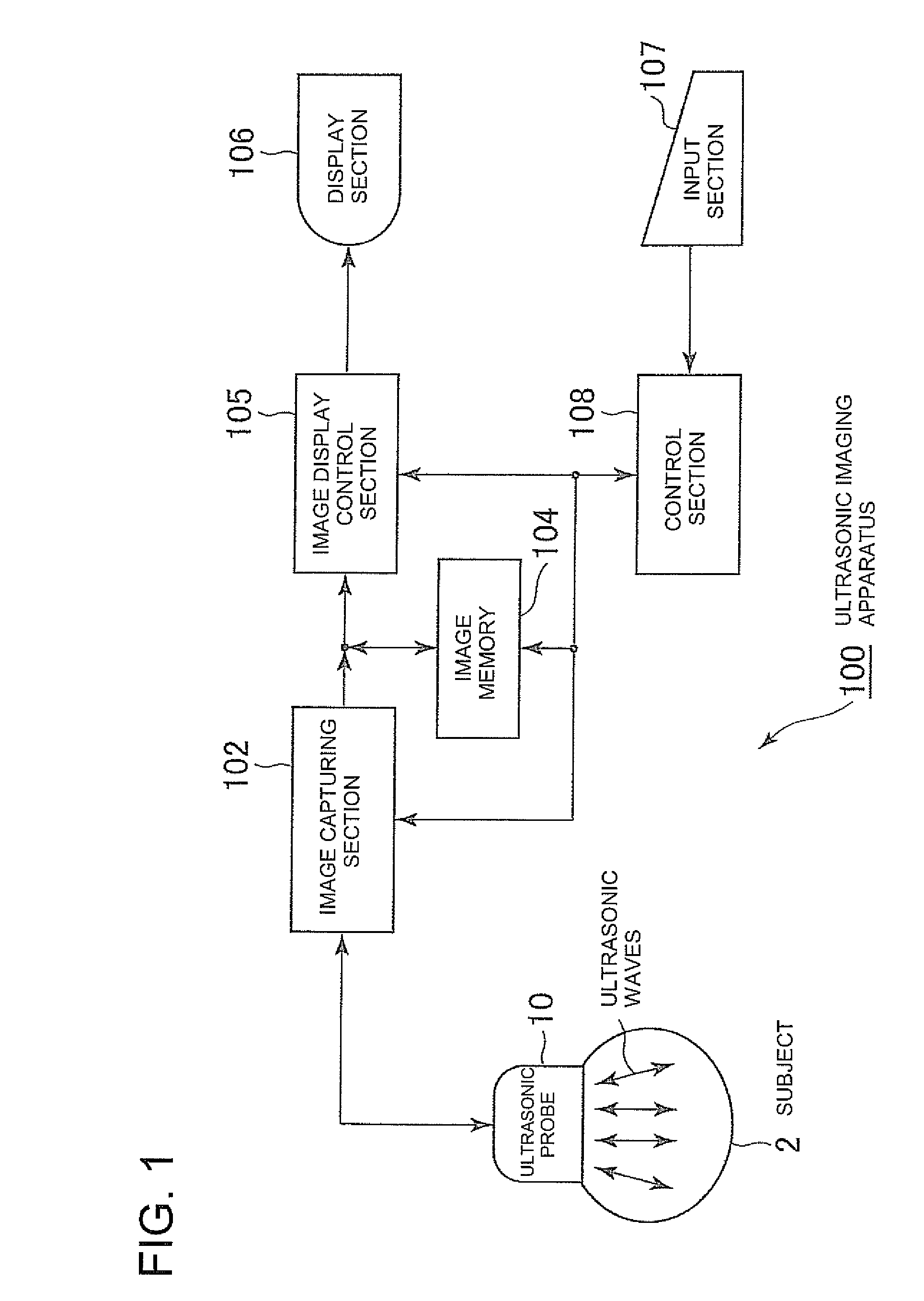
[0055]First, the general structure of an ultrasonic imaging apparatus 100 according to this embodiment will be described. FIG. 1 is a block diagram showing the general structure of the ultrasonic imaging apparatus 100 according to this embodiment. The ultrasonic imaging apparatus 100 includes an ultrasonic probe 10, an image capturing section 102, an image memory 104, an image display control section 105, a display section 106, an input section 107, and a control section 108. Here, the ultrasonic probe 10, the image capturing section 102, the image memory 104 and the control section 108's image capture controller (which will be described later) constitute a 3D tomographic image data capturing means.

[0056]The ultrasonic probe 10 is a part which transmits or receives ultrasonic waves, namely radiates ultrasonic waves in a...


PUM


Abstract

An ultrasonic imaging apparatus includes a 3D tomographic image data capturing device which captures 3D tomographic image data from a 3D region inside a subject, a region-of-interest setting device which sets a 3D region of interest corresponding to the 3D region in an image memory, a surface image extracting device which extracts data on a surface image, in the 3D region of interest, of a massive tissue included in the 3D region, a stereoscopic display generating device which generates stereoscopic display data, and a display device which displays the stereoscopic display data. The region-of-interest setting device displays a 2D tomographic image of a 2D region including the massive tissue on the display device, allows setting of a marker indicating a periphery of the massive tissue in the 2D tomographic image, and generates the 3D region of interest according to data on the marker's position.

Description

CROSS REFERENCE TO RELATED APPLICATIONS[0001]This application claims the benefit of Japanese Patent Application No. 2007-190483 filed Jul. 23, 2007, and incorporated by reference in its entirety.BACKGROUND OF THE INVENTION[0002]The subject matter disclosed herein relates to an ultrasonic imaging apparatus and an image processing apparatus which extract a surface image of a massive tissue in a subject from 3D tomographic image data on the subject and display it stereoscopically.[0003]Recently, in the field of diagnostic imaging using an ultrasonic imaging apparatus, volume rendering in which a surface image of a massive tissue such as a fetus is extracted according to 3D tomographic image data and displayed stereoscopically has been performed. This stereoscopic display makes it easy for an operator to get an overall picture of the massive tissue (for example, see Japanese Patent Application No. 2003-204963).[0004]In this surface image extraction, positional information on a massive t...

Claims


Application Information

Patent Timeline
Patent Type & Authority Applications(United States)
IPC IPC(8): A61B8/00
CPCA61B8/0866A61B8/14A61B8/4461A61B8/483A61B8/463A61B8/469A61B8/461A61B8/466
Inventor KAWAE, SOTARO
Owner GE MEDICAL SYST GLOBAL TECH CO LLC
Why Eureka
Try Eureka