
Apparatus and methods for automated diffusion filtration, culturing and photometric detection and enumeration of microbiological parameters in fluid samples

a technology of microorganisms and fluid samples, applied in biochemistry apparatus, biochemistry apparatus and processes, laboratory apparatus, etc., can solve the problems of high error rate, standard membrane filtration method, and high microbial count of samples, so as to reduce light scattering and control the direction and flow rate

Inactive Publication Date: 2008-11-27
KANIPAYOR RAVI +1
View PDF1 Cites 37 Cited by
  • Summary
  • Abstract
  • Description
  • Claims
  • Application Information

AI Technical Summary

Benefits of technology

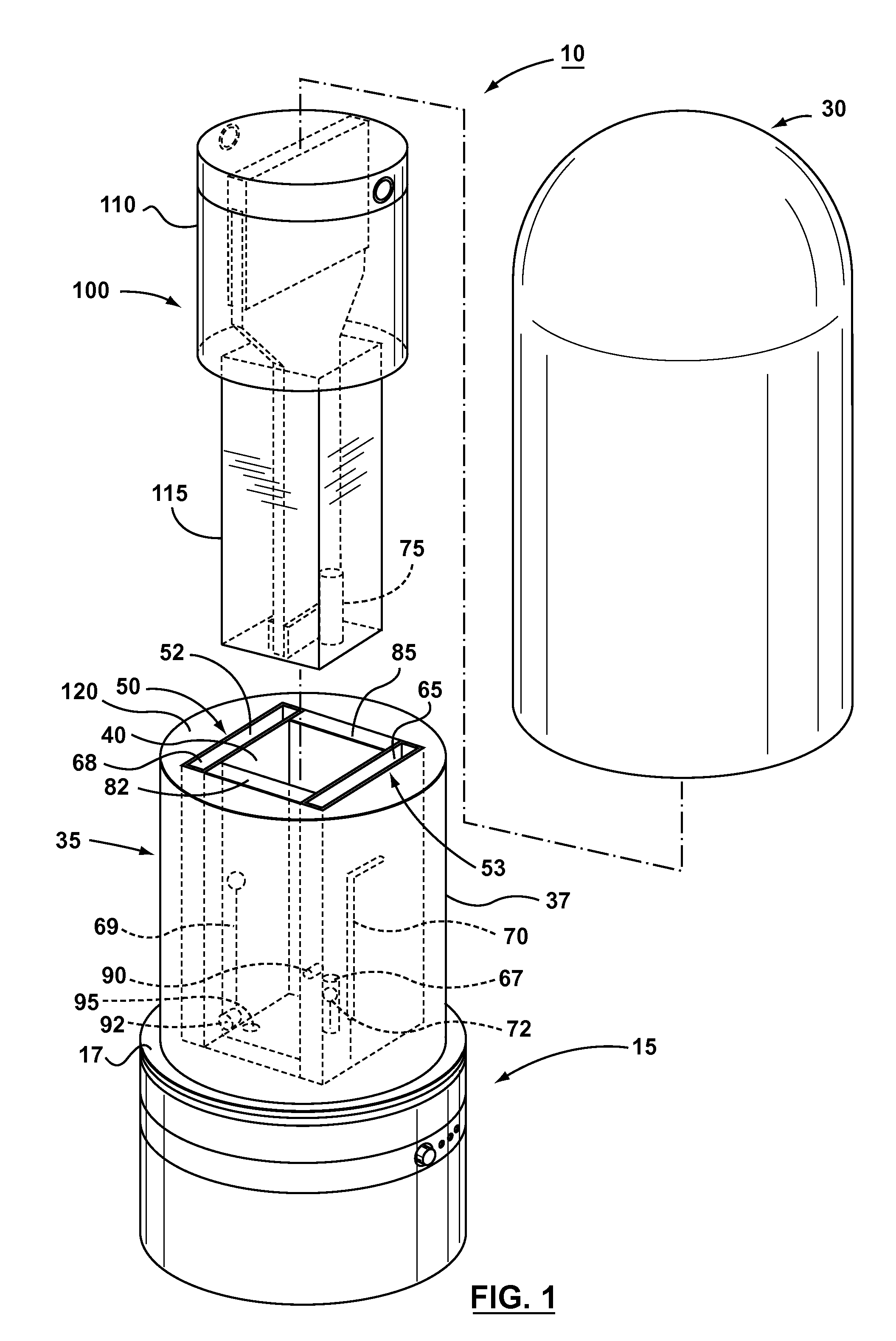
[0009]The sealed specimen cuvette acts as a non-intrusive filter apparatus, a specimen culturing vessel as well as an optical sample cuvette. By connecting the outlet from the detection chamber to a vacuum pump, fluid samples can be conveniently drawn into the culture chamber and through the permeable membrane into the detection chamber until the fluid level reaches the volume mark level in the cuvette. The fluid portion in the detection chamber is filtered according to the pore size of the membrane. The pore size can be suitably selected to retain the microbes under investigation in the culture chamber. The membrane is permeable and thus allows movement of fluid sample along with dissolved components in both directions. By connecting both outlets to a bidirectional pump the direction and rate of the flow can be controlled. As an alternative, a closed circulation loop can be created by connecting both outlets through a peristaltic pump or other suitable devices. This way fluid samples from detection chamber can be drawn back into the culture chamber thus providing homogenization of the dissolved components in both chambers at any given time.
[0010]The specimen cuvette is made of material that allows the propagation of light. The cuvette may be completely cylindrical or square, with planar vertical surfaces, on opposite sides. Flat, planar surfaces reduces light scattering when using photometric detection system.

Problems solved by technology

However, the standard membrane filtration method has several drawbacks.
It is very imprecise, and has problems in dealing with samples containing high microbial counts.
The method is highly error prone due to uncertainty in capturing the target organism within the measurable range.
These photometric methods are very sensitive, and can detect the presence of a very low concentration of color producing components of interest in fluid samples (in parts per million), whereas the human eye can only detect the color when these components are present in very high concentration.
This is not only time consuming but requires separate incubators and spectrometers, and technical personnel to conduct the tests, using robotic sampling systems in some cases.
There is also a potential risk of cross contamination and human error, if proper care is not applied in conducting the analysis.
However, this apparatus is not suitable for use in testing some samples, such as samples containing components that are toxic to the microbiological organism under investigation, and samples containing high suspended solids that can interfere with optical measurements.

Method used


Image

  • Apparatus and methods for automated diffusion filtration, culturing and photometric detection and enumeration of microbiological parameters in fluid samples
  • Apparatus and methods for automated diffusion filtration, culturing and photometric detection and enumeration of microbiological parameters in fluid samples
  • Apparatus and methods for automated diffusion filtration, culturing and photometric detection and enumeration of microbiological parameters in fluid samples

Examples


Embodiment Construction

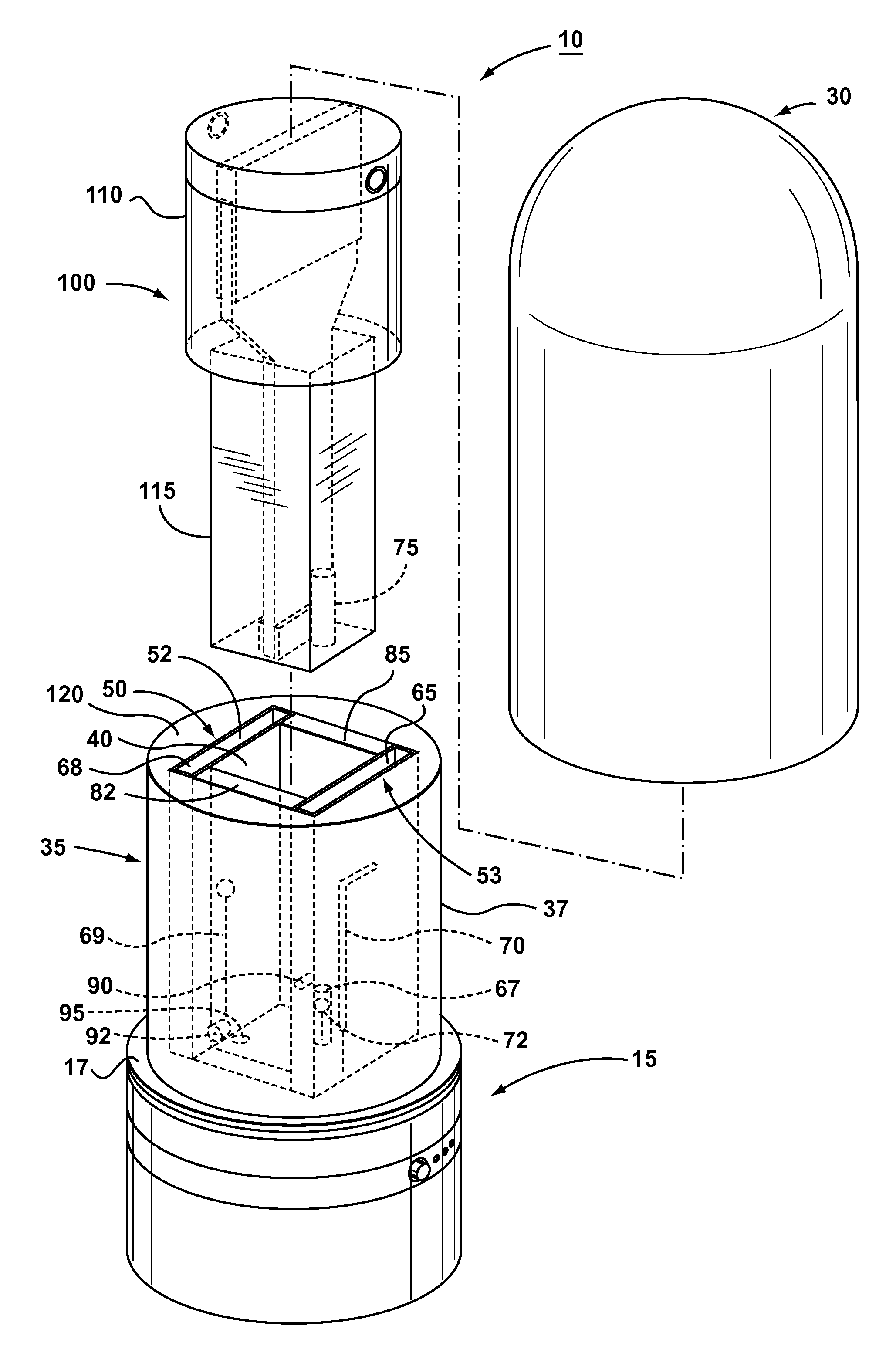
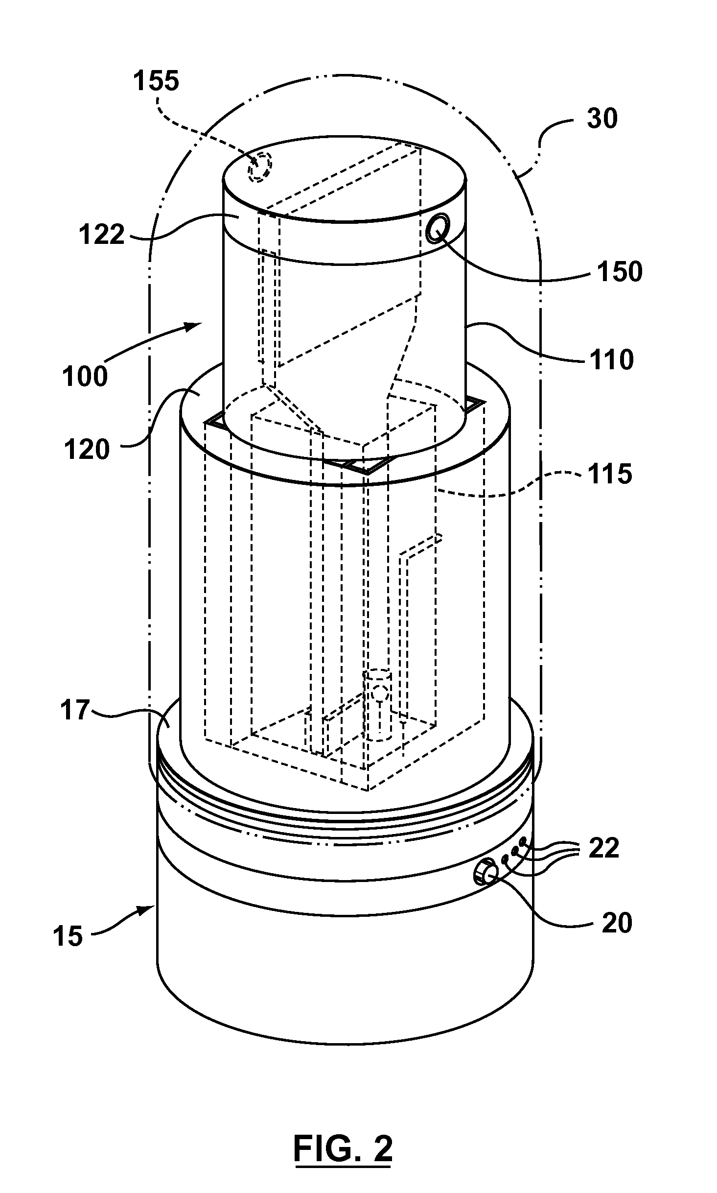
[0022]Referring now to FIGS. 1-6, illustrated therein is an apparatus 10 for photometric detection of microbiological parameters, made in accordance with an embodiment of the present invention. Apparatus 10 comprises a sample cuvette 100, a housing comprising a base unit 15, a cuvette holder 35 mounted on the base unit 15, and a removable lid 30 shaped for enclosing the sample cuvette 100 and the cuvette holder 35. The base unit 15 is generally cylindrical and the cuvette holder 35 extends upwardly from the middle of the top surface 17 of the base unit 15. The cuvette holder 35 has a generally cylindrical outside surface 37, and is provided with a centrally located sample cavity 40 shaped to accommodate the sample cuvette 100. The dimensions of the sample cavity 40 are such that when the sample cuvette 100 is placed in the cuvette holder 35, the sample cavity 40 provides a very snug fit for enhanced photometric detection.

[0023]As best shown in FIGS. 1 and 5, sample cavity 40 is gene...


PUM


Abstract

A system providing non-intrusive, automated culturing and photometric detection for analyzing microbiological parameters is described. The system includes a sealed non-intrusive sample cuvette, a housing with an enclosable cover, systems for incubation and photometric detection mounted within the housing. The sample cuvette consists of a clear graduated optically transmitive container with two chambers—a culture chamber and a detection chamber, separated by a permeable membrane wall. The cuvette has an upper part and a lower part of different dimensions. The upper part is bigger in size than the lower part. The sealed top of the cuvette has two fluid inlet / outlet ports for the introduction of the sample into one chamber while when connected to a suitable vacuum device, the second chamber receives the filtered sample through the permeable membrane. The housing has a cuvette holder that is shaped to provide a very snug fit for the cuvette. When placed inside the cuvette holder the bottom of the upper part of the cuvette rests on top of the holder while the bottom part snugly fits inside the holder cavity. The housing with the enclosable cover provides a thermal chamber during simultaneous incubation and photometric enumeration of microbiological parameters. The cuvette holder accommodates a heating element and a temperature sensor. Photometric detection components comprising LEDs and detectors are placed strategically within the holder.

Description

FIELD OF THE INVENTION[0001]This invention relates to methods and apparatus for the rapid photometric detection and enumeration of microbiological materials in fluid samples.BACKGROUND OF THE INVENTION[0002]The membrane filtration method is the standard for testing microbiological parameters in water samples. One advantage of the standard membrane filtration method is that it separates the organism under investigation from the potential toxic components present in the original sample matrix. However, the standard membrane filtration method has several drawbacks. The method is over 200 years old and no major modifications have been done to improve the methodology. It is very imprecise, and has problems in dealing with samples containing high microbial counts. The standard membrane filtration method requires manual, visual counting of the growth on the filter as well as experience in identifying false growth. The method is highly error prone due to uncertainty in capturing the target ...

Claims


Application Information

Patent Timeline
IPC IPC(8): C12Q1/02C12M1/34C12M1/36C12M1/00
CPCC12M23/12C12M23/34C12M29/04C12M41/36B01L3/502B01L2300/0681B01L2300/0809B01L2300/0832B01L2300/0858
Inventor KANIPAYOR, RAVIEMBURGH, RON
Owner KANIPAYOR RAVI
Learn More