
Systems and methods for calibrating functional magnetic resonance imaging of living tissue

a functional magnetic resonance imaging and living tissue technology, applied in the field of systems and methods for calibrating functional magnetic resonance imaging of living tissue, can solve the problems of difficult to establish a calibration standard for in vivo fmri data, affecting the repeatability of acquiring bold/boss contrast signals, and challenging calibration within the field of fmri

Inactive Publication Date: 2008-11-13
UNIV OF FLORIDA RES FOUNDATION INC
View PDF14 Cites 7 Cited by
  • Summary
  • Abstract
  • Description
  • Claims
  • Application Information

AI Technical Summary

Benefits of technology

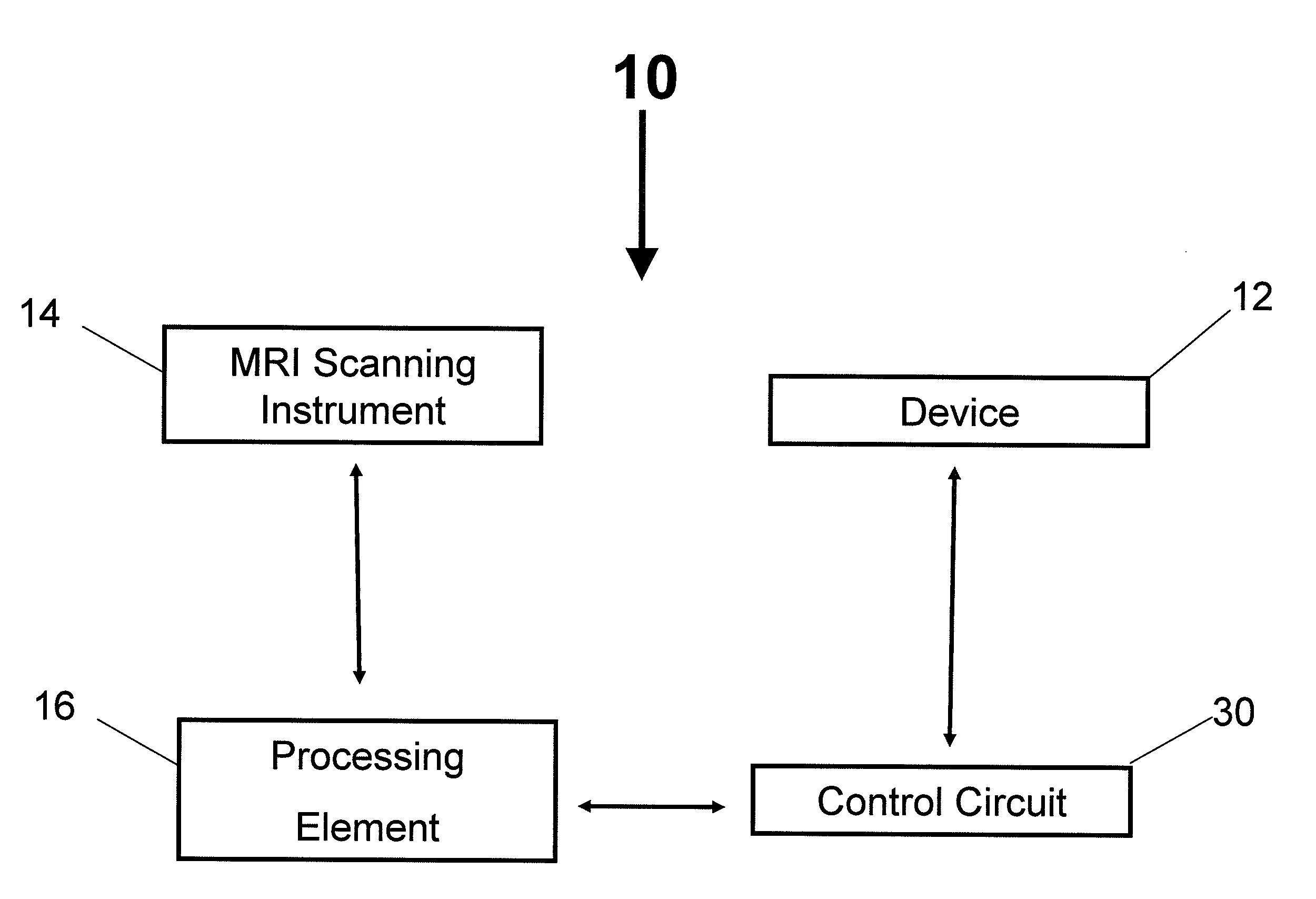
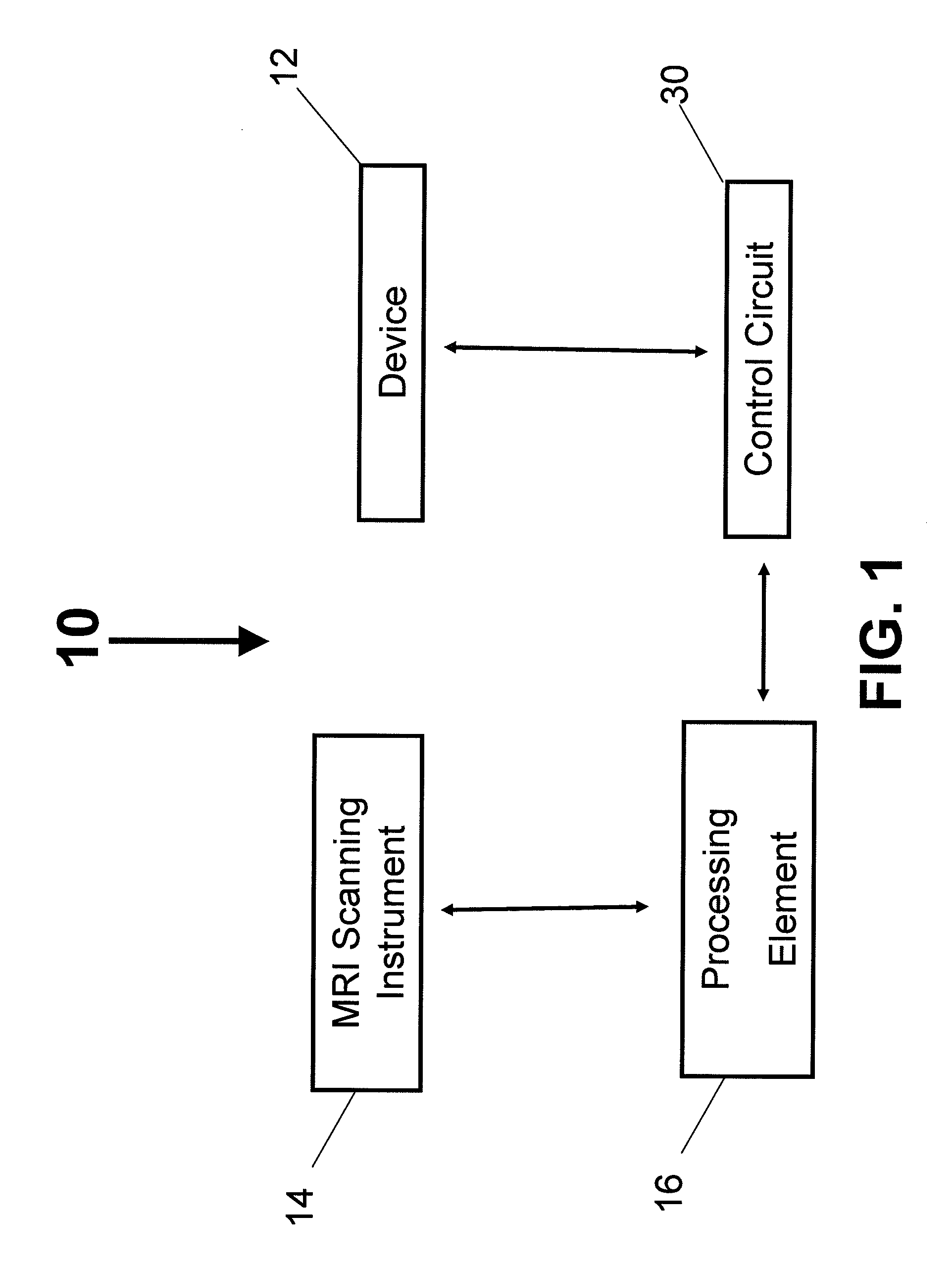
[0008]Embodiments of the present invention relate to systems and methods for calibrating functional magnetic resonance imaging of living tissue. For example, a calibration method includes providing a contrast signal simulation device configured for generating one or more pre-determined BOLD and/or BOSS contrast simulation signals that simulate at least one BOLD/BOSS contrast signal generated by living tissue of a subject (e.g., a human being or a living animal) and positioning the contrast signal simulation device adjacent to the subject. The method also includes using an imaging instrument (e.g., a magnetic resonance imaging device) to take a first data set from said subject's tissue while: (A) the contrast signal simulation device is disposed adjacent to the subject, and (B) the contrast signal simulation device is generating the one or more pre-determined BOLD/BOSS contrast simulation signals. The method further includes using the imaging instrument to take a second data set from the subject's tissue while the contrast signal simulation device is not generating the one or more pre-determined BOLD/BOSS contrast simulation signals and using the first and second data sets to determine at least one figure of merit of the imaging instrument, the contrast simulation device, and/or the living tissue (e.g., a sensitivity, a specificity, and/or a receiver operating characteristic).
[0009]According to aspects of the method, the figure of merit of the imaging instrument takes into account in vivo noise associated with the subject. In addition, the step of using the first and second data sets to determine at least one figure of merit may include determining statistically reliable differences between the first and second data sets. The method may further include using the imaging instrument to acquire a third data set of the subject's tissue while the subject is being subjected to a particular task or event and using the imaging instrument to take a fourth data set of the subject's tissue while the subject is not being subjected to the particular task or event. In addition, the method may

Problems solved by technology

Calibration within the field of fMRI is challenging because repeatability is imperfect—even when using the same procedures with the same subject.
It is difficult to establish a calibration standard for in vivo fMRI data.
In particular, the presence of instrument noise and “physiological noise” produced by a living human brain (or by extension, produced by other living tissue) affects the repeatability of acquiring BOLD / BOSS contrast signals.
Thus, current techniques for calibrating the sensitivity and specificity of an fMRI test for detecting BOLD / BOSS contrast signals in the presence of such variable and idiosyncratic noise remain inadequate.

Method used


Image

  • Systems and methods for calibrating functional magnetic resonance imaging of living tissue
  • Systems and methods for calibrating functional magnetic resonance imaging of living tissue
  • Systems and methods for calibrating functional magnetic resonance imaging of living tissue

Examples


Embodiment Construction

[0014]The present invention will now be described more fully hereinafter with reference to the accompanying drawings, in which various embodiments of the invention are shown. This invention may, however, be embodied in many different forms and should not be construed as limited to the embodiments set forth herein. Rather, these embodiments are provided so that this disclosure will be thorough and complete, and will fully convey the scope of the invention to those skilled in the art. Like numbers refer to like elements throughout.

Overview

[0015]A method of calibrating an fMRI instrument according to a particular embodiment of the present invention comprises the steps of: (1) providing a BOLD / BOSS contrast signal simulation device that is configured for generating one or more pre-determined BOLD and / or BOSS contrast simulation signals that serve to simulate one or more endogenous BOLD / BOSS contrast signals generated by living tissue (e.g., a human brain); (2) positioning the BOLD / BOSS ...


PUM


Abstract

Embodiments of the present invention relate to systems and methods for calibrating functional magnetic resonance imaging of living tissue. For example, a calibration method includes providing a contrast signal simulation device configured for generating one or more pre-determined contrast simulation signals selected from a group consisting of BOLD and BOSS contrast simulation signals that simulate at least one contrast signal generated by living tissue of a subject and positioning the contrast signal simulation device adjacent to the subject. The method includes using an imaging instrument to take first and second data sets from the subject's tissue while the contrast signal simulation device is disposed adjacent to the subject and while generating and not generating the one or more pre-determined contrast simulation signals, respectively. The method also includes using the first and second data sets to determine at least one figure of merit of the imaging instrument, the contrast simulation device, and / or the living tissue.

Description

CROSS-REFERENCE TO RELATED APPLICATIONS[0001]The present application is a Continuation of International Application No. PCT / US2007 / 066869, filed Apr. 18, 2007, which claims the benefit of U.S. Provisional Application No. 60 / 793,210, filed Apr. 19, 2006. The disclosures of both referenced applications are incorporated herein by reference.BACKGROUND OF THE INVENTION[0002]Functional Magnetic Resonance Imaging, or fMRI, includes imaging techniques that may be used to determine which areas of a patient's brain (or other tissues) are activated by different types of activities, such as the patient moving their fingers or reacting to a particular image. To execute an fMRI scan of the brain, for example, an MRI machine is first configured to image increased blood flow to activated brain areas. A task event is then provided that is likely to cause activity, and changes in blood flow, in the patient's brain. (Such events may include, for example, viewing pictures that may be selectively presen...

Claims


Application Information

Patent Timeline
IPC IPC(8): A61B5/055
CPCG01R33/4806G01R33/58
Inventor WHITE, KEITH DAVID
Owner UNIV OF FLORIDA RES FOUNDATION INC
Learn More