
Response Predictors for Erbb Pathway-Specific Drugs

a technology of erbb pathway and response predictor, which is applied in the field of response predictors for erbb pathway specific drugs, can solve the problems of disease progression, egfr expression by tumor cells, and individual receptor expression level alone is not always a reliable indicator, and achieves positive or negative correlation with cell or tissue responsiveness

Inactive Publication Date: 2008-10-16
MONOGRAM BIOSCIENCES
View PDF0 Cites 41 Cited by
  • Summary
  • Abstract
  • Description
  • Claims
  • Application Information

AI Technical Summary

Benefits of technology

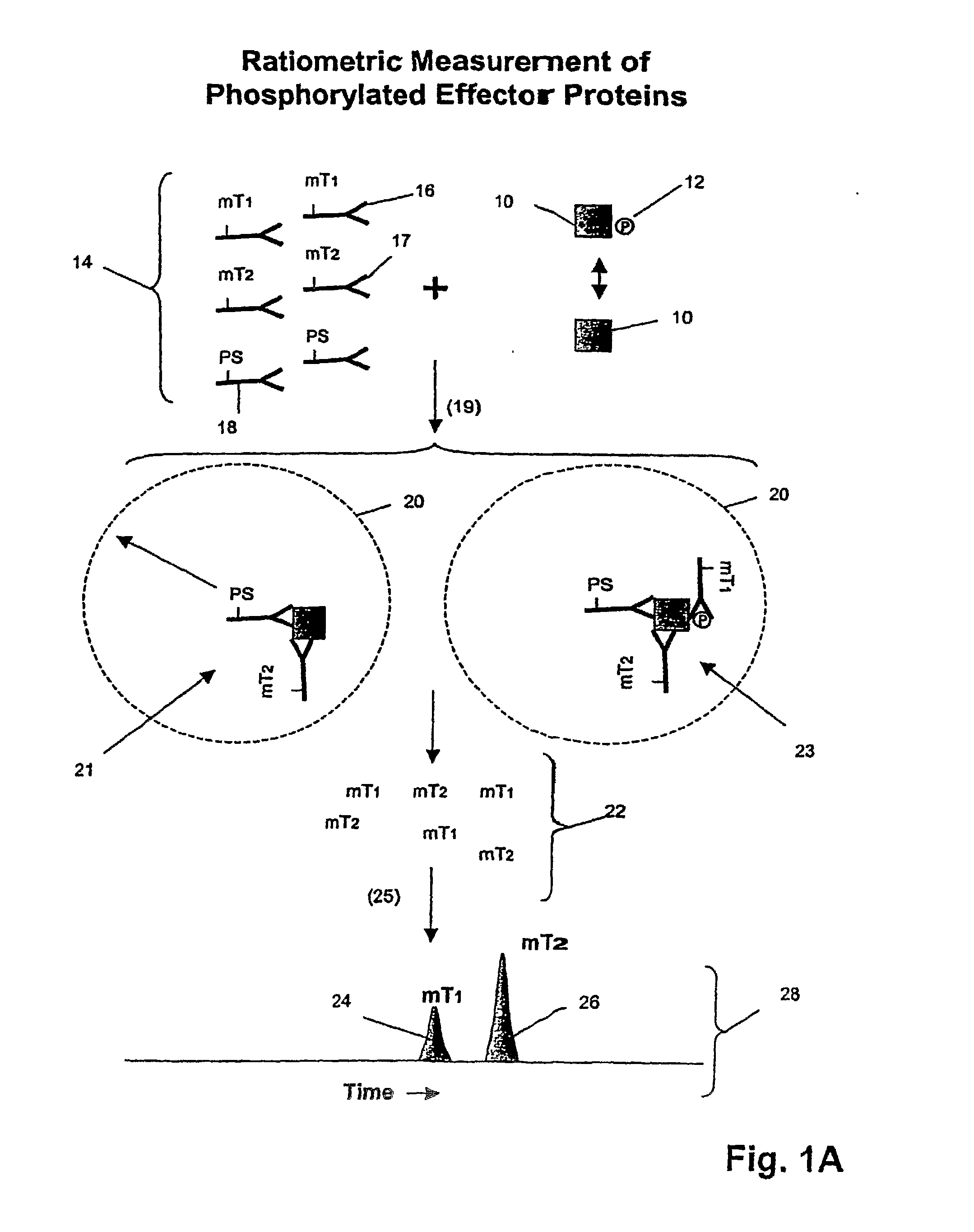
[0011]In one aspect, the invention provides a method of determining whether tumor cells or tissue is responsive to treatment with an ErbB pathway-specific drug. In accordance with this aspect, measurements are made on such cells or tissues to determine values for total ErbB receptors of one or more types, ErbB receptor dimers of one or more types and their phosphorylation states, and/or one or

Problems solved by technology

However, individual receptor expression level alone is not always a reliable indicator of a disease status or condition, e.g. Chow et al, Clin.
Despite the important role that receptor dimerization plays in cellular and disease processes, receptor dimer expression has not been employed as a biomarker, in part due to the inconvenience and lack of sensitivity of current measurement technologies and the inability or impracticality of using such technologies to carry out measurements on patient samples, which may be formalin fixed and / or in too small a quantity for analysis, e.g. Price et al, Methods in Molecular Biology, 218: 255-267 (2003); Stagljar, Science STKE 2003, pe56 (2003); Koll et al, International patent publication WO 2004 / 008099; Golemis, editor, Protein-Protein Interactions (Cold Spring Harbor Laboratory Press, New York, 2002); Sorkin et al, Curr. Biol., 10: 1395-1398 (2000); McVey et al, J. Biol. Chem., 17: 14092-14099 (2001); Salim et al, J. Biol. Chem., 277: 15482-15485 (2002); Angers et al, Annu. Rev. Pharmacol. Toxicol., 42: 409-435 (2002); Szollosi et al, Reviews in Molecular Biotechnology, 82: 251-266 (2002); Matko et al, Meth. in Enzymol., 278: 444-462 (1997); Reed-Gitomer, U.S. Pat. No. 5,192,660.
EGFR expression by tumor cells has been linked with aggressive tumor growth, disease progression, poor survival, and poor response to therapy.
In a similar manner, ErbB2 has been reported to be over expressed in 15-30% of invasive ductal breast cancer and has been associated with increased proliferation, poor clinical outcome, and altered responsiveness to various adjuvant therapies.

Method used


Image

  • Response Predictors for Erbb Pathway-Specific Drugs
  • Response Predictors for Erbb Pathway-Specific Drugs
  • Response Predictors for Erbb Pathway-Specific Drugs

Examples


example 1

7.2 Example 1

Simultaneous Measurement of Her2-Her3 Heterodimerization and Erk1 Phosphorylation

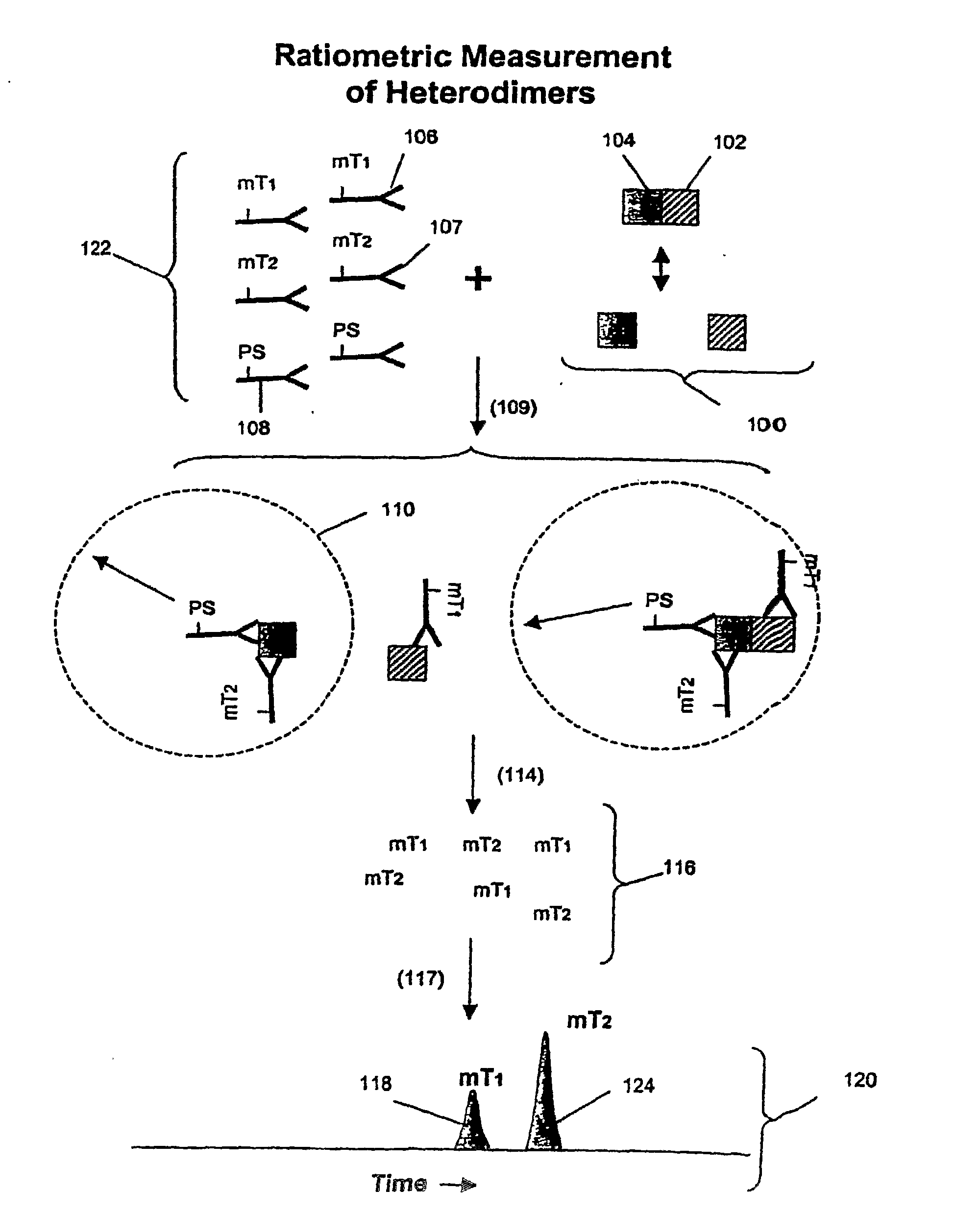
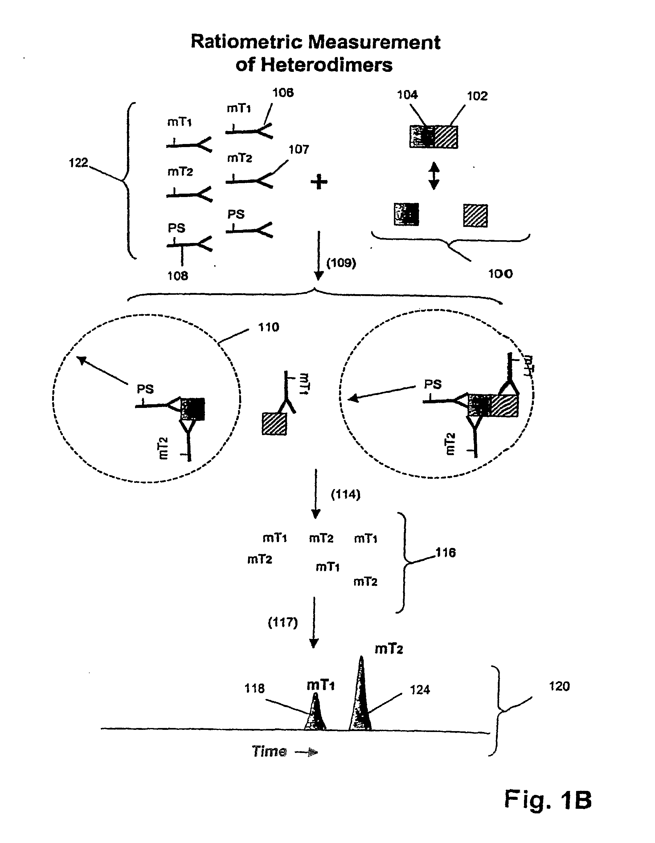
[0181]In this example, an assay is described for providing a ratiometric measure of phosphorylated Erk1 and Her2-Her3 heterodimerization. The assays are carried out as follows.

[0182]Sample Preparation:[0183]1. Serum-starve breast cancer cell line culture (MCF-7) overnight before use.[0184]2. Stimulate cell lines with HRG in culture media for 10 minutes at 37° C. Exemplary doses of HRG are 0, 0.032, 0.16, 0.8, 4, 20, 100 nM for MCF-7 cells.[0185]3. Aspirate culture media, transfer onto ice, and add lysis buffer (described below) to lyse cells in situ.[0186]4. Scrape and transfer lysate to microfuge tube. Incubate on ice for 30 min. Microfuge at 14,000 rpm, 4° C., for 10 min.[0187]5. Collect supernatants as lysates and aliquot for storage at −80° C. until use.

Lysis Buffer (Made Fresh and Stored on Ice):

[0188]

FinalulStock1% Triton X-100100010%20 mM Tris-HCl (pH 7.5)200  1 M100 mM NaCl200  5 M5...

example 2

7.3 Example 2

Analysis of Cell Lysates for Her-2 Heterodimerization and Receptor Phosphorylation

[0207]In this example, Her1-Her2 and Her2-Her3 heterodimers and phosphorylation states are measured in cell lysates from several cell lines after treatment with various concentrations of epidermal growth factor (EGF) and heregulin (HRG). Measurements are made using three binding compounds and a cleaving probe as described below.

[0208]7.3.1 Sample Preparation:[0209]1. Serum-starve breast cancer cell line culture overnight before use.[0210]2. Stimulate cell lines with EGF and / or HRG in culture media for 10 minutes at 37° C. Exemplary doses of EGF / HRG are 0, 0.032, 0.16, 0.8, 4, 20, 100 DM for all cell lines (e.g. MCF-7, T47D, SKBR-3) except BT20 for which the maximal dose is increased to 500 nM because saturation is not achieved with 100 nM EGF.[0211]3. Aspirate culture media, transfer onto ice, and add lysis buffer to lyse cells in situ.[0212]4. Scrape and transfer lysate to microfuge tube....

example 3

7.4 Example 3

Analysis of Tissue Lysates for Her2 Heterodimerization and Receptor Phosphorylation

[0240]In this example, Her1-Her2 and Her2-Her3 heterodimers and phosphorylation states are measured in tissue lysates from human breast cancer specimens.

[0241]7.4.1 Sample Preparation:[0242]1. Snap frozen tissues are mechanically disrupted at the frozen state by cutting.[0243]2. Transfer tissues to microfuge tube and add 3× tissue volumes of lysis buffer (from appendix I) followed by vortexing to disperse tissues in buffer.[0244]3. Incubate on ice for 30 min with intermittent vortexing to mix.[0245]4. Centrifuge at 14,000 rpm, 4° C., for 20 min.[0246]5. Collect supernatants as lysates and determine total protein concentration with BCA assay (Pierce) using a small aliquot.[0247]6. Aliquot the rest for storage at −80° C. until use.

[0248]7.4.2 Assay Design:[0249]1. The total assay volume is 40 ul.[0250]2. The lysates are tested in serial titration series of 40, 20, 10, 5, 2.5, 1.25, 0.63, 0....


PUM


Abstract

The invention provides a method of determining whether tumor cells or tissue is responsive to treatment with an ErbB pathway-specific drug. In accordance with the invention, measurements are made on such cells or tissues to determine values for total ErbB receptors of one or more types, ErbB receptor dimers of one or more types and their phosphorylation states, and / or one or more ErbB signaling pathway effector proteins and their phosphorylation states. These quantities, or a response index based on them, are positively or negatively correlated with cell or tissue responsiveness to treatment with an ErbB pathway-specific drug. In one aspect, such correlations are determined from a model of the mechanism of action of a ErbB pathway-specific drug on an ErbB pathway. Preferably, methods of the invention are implemented by using sets of binding compounds having releasable molecular tags that are specific for multiple components of one or more complexes formed in ErbB pathway activation. After binding, molecular tags are released and separated from the assay mixture for analysis.

Description

1. FIELD OF THE INVENTION[0001]The present invention relates generally to methods for determining the status of ErbB receptors of a cell or tissue, and more particularly, for using such status information to select patients responsive to ErbB receptor-specific drugs.2. BACKGROUND OF THE INVENTION[0002]A biomarker is a characteristic that is objectively measured and evaluated as an indicator of normal biological processes, pathogenic processes, or pharmacological responses to a therapeutic intervention, Atkinson et al, Clin. Pharmacol. Ther., 69: 89-95 (2001). Biomarkers vary widely in nature, ease of measurement, and correlation with physiological states of interest, e.g. Frank et al, Nature Reviews Drug Discovery, 2: 566-580 (2003). It is widely believed that the development of new validated biomarkers will lead both to significant reductions in healthcare and drug development costs and to significant improvements in treatment for a wide variety of diseases and conditions. Thus, a ...

Claims


Application Information

Patent Timeline
IPC IPC(8): C12Q1/02
CPCG01N33/57492G01N2800/52
Inventor SINGH, SHARAT
Owner MONOGRAM BIOSCIENCES
Learn More