
Assay for porcine circovirus production

a technology of porcine circovirus and porcine intestine, which is applied in the field of porcine circovirus production, can solve the problems of insufficient treatment and prevention of this disease, and treatment must be performed with caution

Active Publication Date: 2008-10-02
MERIAL INC
View PDF0 Cites 6 Cited by
  • Summary
  • Abstract
  • Description
  • Claims
  • Application Information

AI Technical Summary

Benefits of technology

This method enables rapid and efficient monitoring of viral production, allowing for the selection of an optimal harvesting point that achieves high viral yields suitable for vaccine production, reducing the incubation period and associated costs, while providing quantitative data for repeated monitoring throughout the incubation period.

Problems solved by technology

Treatment and prevention of this disease are not currently available.
However, the treatment must be performed with caution because D-glucosamine is toxic for cell culture (see, Allan et al.

Method used


Image

  • Assay for porcine circovirus production
  • Assay for porcine circovirus production
  • Assay for porcine circovirus production

Examples


example 1

Correspondence between the Designations of ORFs of Circovirus PCV2

[0081]The ORFs of circovirus PCV2, their equivalent designators, and the respective sources thereof, incorporated herein by reference in the entirety, are shown in Table 1 below. ORF1 and ORF2, as described by Meehan et al. (1997; 1998) have been alternatively designated as ORFs 4 and 13 respectively.

TABLE 1PCV2 ORF numbering and equivalentsExample 2: Assay of viability, Detecting ORF1 andORF2 in PCV2-infected PK-15 cellsORF NumberingAlternative DesignationsMeehan et al.U.S. patentU.S. Patent Ser. Nos.J. Gen. Virol.application Ser.6,368,601, 6,391,314,78: 221-227No. 200201066396,660272, 6,217,883,Source(1997)to Wang et al.6,517,843, 6,497,8831142613327431054565371829610811912111312

(a) Determination of Cell Counts and Cell Viability

[0082]Propidium iodide was used to assess plasma membrane integrity. Propidium iodide is a fluorescent vital dye that stains nucleic acid. Dead cells incorporate propidium iodide which was d...

example 3

PK-15 Count and Viability after PCV2 Inoculation in Batch Seed Cultures

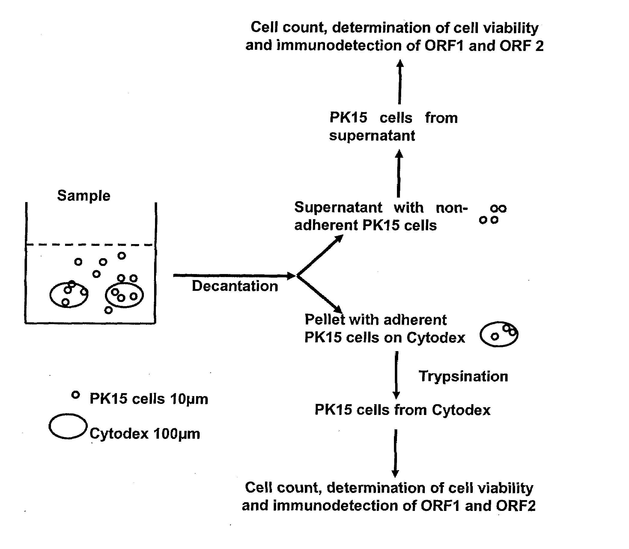
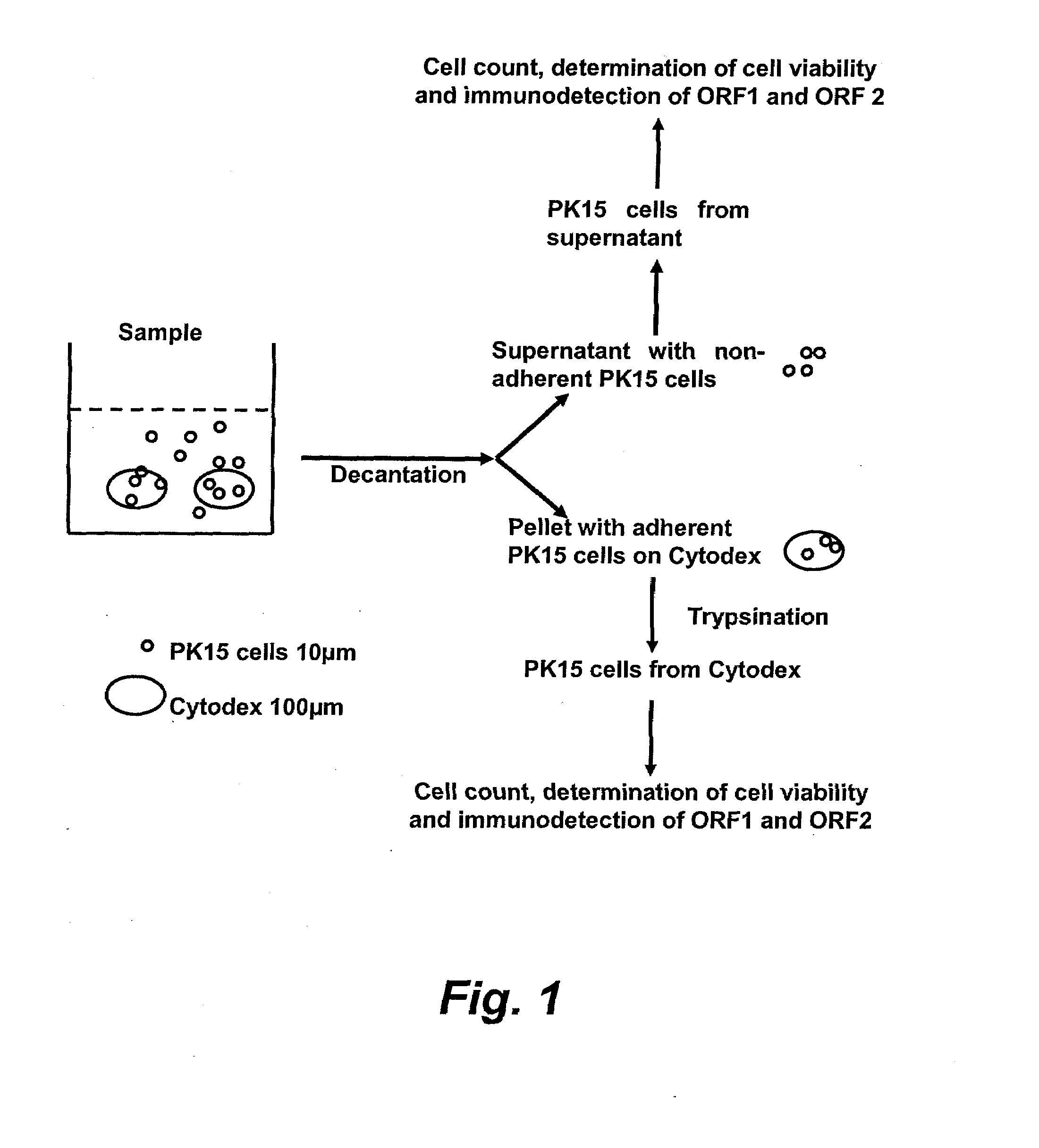
[0088]As shown in FIG. 3, At day 5 of the incubation period of a batch seed culture of PCV2 virus growing on PK-15 cells, 50% of the host cells were located in the supernatant and were mostly dead (73%). In contrast, 50% of the host cells were located on the CYTODEX™ (Amersham Biosciences, Inc) substratum support and were almost entirely a viable population (96%).

[0089]PCV2-infected PK-15 cells were detected at the end of the culture at day 5, but only in the supernatant cells using flow cytometry to detect ORF1 and ORF2. As shown, for example, in FIG. 6, the kinetics of the viral-specific antigen formation was dependent on the ORF: ORF1 was produced early and at low levels whilst ORF was produced later in the incubation cycle and at increasing levels.

example 4

ORF1 and ORF2 Total Staining

[0090]As shown in FIG. 6, for ORF1 there was no signal on cells membrane detected during the later phase of the culture incubation period. Table 2, below, shows that no membrane signals were detected on cells adherent to CYTODEX™ (Amersham Biosciences, Inc). Major ORF-specific signals were on cells from the culture supernatant and were increasing from day 2 to day 5. Results were similar for live or dead cells, with a maximum of 60% at day 4.

TABLE 2Count(total / ml)Cells from supernatantAdherent cells% of3.6 × 105Membrane +4.7 × 10⁵Membrane +Total Cells43%Intracellular57%IntracellularStainingMembranecompartments.Membranecompartments.ORF135%ORF2  37% 60%


PUM


Abstract

The present invention provides methods for the determination of the viral titer of a culture of host animal host cells infected with a circovirus. The FACS-based methods of the invention may include determining the viability of the host cells in a cell culture medium supernatant and of those cells that remain adherent to a solid support. Detecting and measuring the percentage of cells that expressed the viral antigens ORF1 and ORF2 may determine the viral load of the cultured host cells. The yield of the virus may be established by the detection and measurement of both antigens in supernatant cells, for example 5 to 7 days from when the host cells are transferred to a serum free medium. The methods of the invention may yield rapid quantitative data. This allows the repeated in-process monitoring of the viral production throughout the incubation period, and ready selection of the most appropriate harvesting point.

Description

RELATED APPLICATIONS[0001]This application claims priority to Provisional U.S. Application Ser. No. 60 / 670,892 filed Apr. 13, 2005, the contents of which are hereby expressly incorporated herein by reference.INCORPORATION BY REFERENCE[0002]All documents cited therein or during their prosecution (“application cited documents”) and all documents cited or referenced in the application cited documents, and all documents cited or referenced herein (“herein cited documents”), and all documents cited or referenced in herein cited documents, together with any manufacturer's instructions, descriptions, product specifications, and product sheets for any products mentioned herein or in any document incorporated by reference herein, are hereby incorporated herein by reference, and may be employed in the practice of the inventionFIELD OF THE INVENTION[0003]This invention relates to methods of in-process monitoring of a viral yield during batch incubation production processes using fluorescent an...

Claims


Application Information

Patent Timeline
Patent Type & Authority Applications(United States)
IPC IPC(8): C12Q1/70G01N33/566
CPCA61K2039/525C12N7/00G01N2333/01G01N33/56983C12N2750/10051C12N7/02
Inventor ALLIBERT, PATRICECUPILLARD, LIONEL PIERRE CHRISTIANREYES, JEAN
Owner MERIAL INC
Learn More