
Image-guided delivery of therapeutic tools duing minimally invasive surgeries and interventions

a technology of ultrasound imaging and therapeutic tools, applied in the field of medical devices, can solve the problems of limited vision of existing minimally invasive techniques, difficulty in optical imaging, intravascular or endoscopic ultrasound imaging devices, etc., and achieve the effect of improving imaging

Inactive Publication Date: 2008-09-11
THE BOARD OF TRUSTEES OF THE LELAND STANFORD JUNIOR UNIV
View PDF30 Cites 157 Cited by
  • Summary
  • Abstract
  • Description
  • Claims
  • Application Information

AI Technical Summary

Benefits of technology

[0010]Minimally invasive interventions can be performed by inserting the image-guided therapy device into a body lumen or cavity, imaging a region inside the body lumen by using the annular ultrasound array, guiding the therapeutic tool to a focus spot based on the imaging, and applying therapy to the focus spot by using the therapeutic tool. The interventions can also include scanning over an area inside the body lumen or delivering an ultrasound contrast agent through the inner lumen of the elongate tubular member to enhance the imaging.

Problems solved by technology

However, existing minimally invasive techniques suffer from restricted vision.
Particularly, for cardiac or vascular interventions, blood poses difficulties for optical imaging.
Existing intravascular or endoscopic ultrasound imaging devices, however, typically only provide side-looking cross-sectional images.
For example, in coronary catheterization, totally occluded or heavily stenosed vessels make it impossible to introduce catheters with strictly side-looking capabilities.
Many existing minimally invasive medical instruments are also typically limited to image only or therapy only capabilities.
In the former scenario, the accuracy and guidance capability would be limited and in the latter scenario, the size of the openings must be large enough to accommodate multiple tubes.
However, existing low frequency HIFU devices typically do not offer sufficient focal intensity for tissue ablation, especially for HIFU transducers with small diameters.

Method used


Image

  • Image-guided delivery of therapeutic tools duing minimally invasive surgeries and interventions
  • Image-guided delivery of therapeutic tools duing minimally invasive surgeries and interventions
  • Image-guided delivery of therapeutic tools duing minimally invasive surgeries and interventions

Examples


Embodiment Construction

[0022]Minimally invasive surgeries and interventions require delivery of a therapeutic tool through natural body openings or small artificial incisions. However, many instruments and methods to conduct these interventions suffer from restricted vision. Below is a detailed description of methods and devices for image-guided therapy delivery usable in minimally invasive surgeries and interventions.

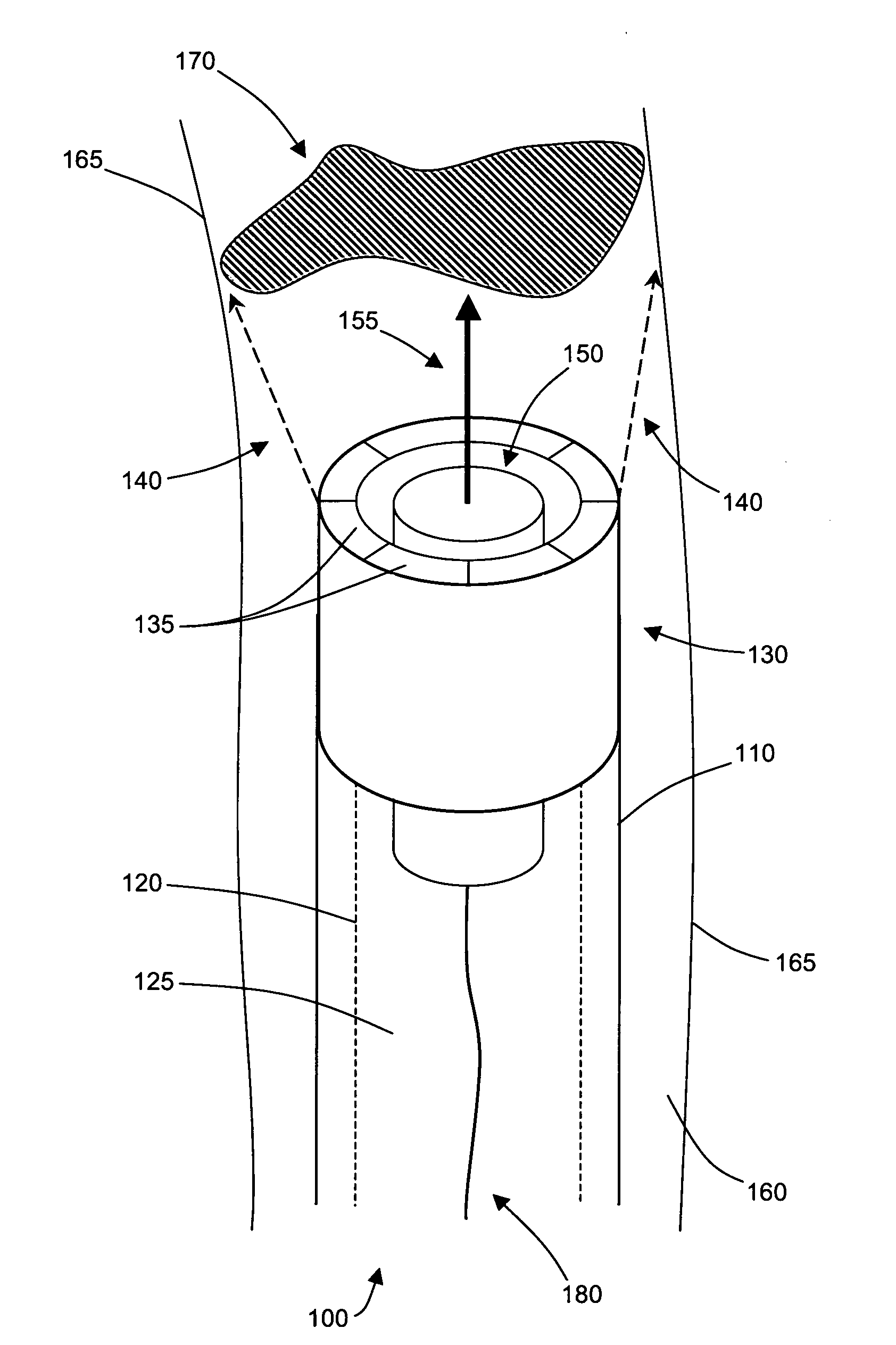
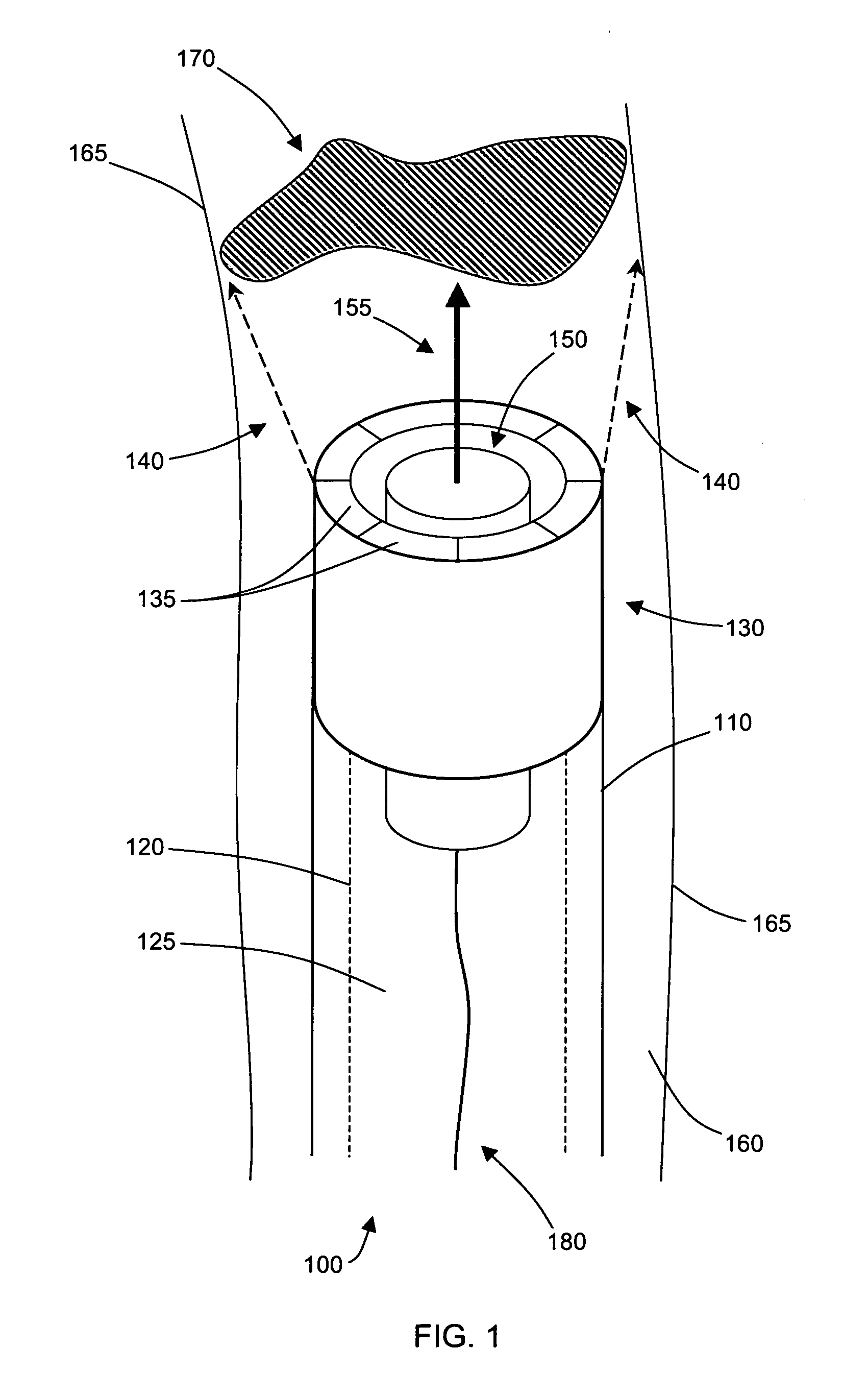
[0023]FIG. 1 shows an example of an image-guided therapy device 100 that has been inserted inside of a body lumen 160. The image-guided therapy device 100 includes an elongate member, such as a catheter or an endoscopic instrument, dimensioned to fit inside of the body lumen 160. The elongate member is tubular and has an outer wall 110 and an inner wall 120. The inner wall 120 forms the inner lumen 125 of the elongate tubular member.

[0024]Located at the distal end of the elongate tubular member are the acoustic imaging and therapy components of the image-guided therapy device 100. An annular...


PUM


Abstract

Imaged-guided therapy for minimally invasive surgeries and interventions is provided. An image-guided device includes an elongate tubular member, such as a catheter, an annular array of capacitive micromachined ultrasound transducers (cMUTs) for real-time three-dimensional forward-looking acoustic imaging, and a therapeutic tool. The therapeutic tool is positioned inside an inner lumen of the elongate tubular member and can be a device for tissue ablation, such as a high intensity focused ultrasound (HIFU) device or a laser. The HIFU device is operable at high frequencies to have a sufficiently small focus spot, thus a high focal intensity. The imaging annular array is also operable at high frequencies for good acoustic imaging resolution. The high resolution forward-looking imaging array, in combination with the high frequency HIFU transducer, provides a single image-guided therapy device for precise tissue ablation and real-time imaging feedback.

Description

CROSS-REFERENCE TO RELATED APPLICATIONS[0001]This application claims priority from U.S. Provisional Patent Application 60 / 906,097 filed Mar. 7, 2007, which is incorporated herein by reference.STATEMENT OF GOVERNMENT SPONSORED SUPPORT[0002]This invention was made with Government support under contract NIH awarded by 1-RO1-HL7647 and grant number GPEDC0013B from OHSU. The Government has certain rights in the invention.FIELD OF THE INVENTION[0003]The invention relates generally to medical devices. More particularly, the present invention relates to ultrasound image-guided delivery of therapeutic tools for minimally invasive medical procedures.BACKGROUND[0004]Minimally invasive techniques are widely used in medical procedures including cardiac, vascular, joint, abdominal, and spinal surgeries and interventions. In minimally invasive interventions, a surgical or medical tool is introduced into the body through a natural body opening or small artificial incisions. A separate endoscopic ca...

Claims


Application Information

Patent Timeline
IPC IPC(8): A61B8/00A61B18/20A61N7/00
CPCA61B8/0833A61B8/0841A61B8/12A61B8/4483A61B8/4488A61B8/483B06B1/0292A61B18/24A61B19/52A61B2019/5278A61B2562/028A61N7/022A61B10/04A61B90/36A61B2090/3782
Inventor KHURI-YAKUB, BUTRUS T.ORALKAN, OMERYARALIOGLU, GOKSEN G.
Owner THE BOARD OF TRUSTEES OF THE LELAND STANFORD JUNIOR UNIV
Learn More