
Phantom for ion range detection

a technology of ion range and phantom, which is applied in the field of radiotherapy systems using heavy ions, can solve the problems of limited value of portal imaging systems, relying on radiation exiting the patient, and heavy ions that are not easily characterized by entrance dose monitors and portal imaging devices

Active Publication Date: 2008-09-11
WISCONSIN ALUMNI RES FOUND
View PDF56 Cites 23 Cited by
  • Summary
  • Abstract
  • Description
  • Claims
  • Application Information

AI Technical Summary

Benefits of technology

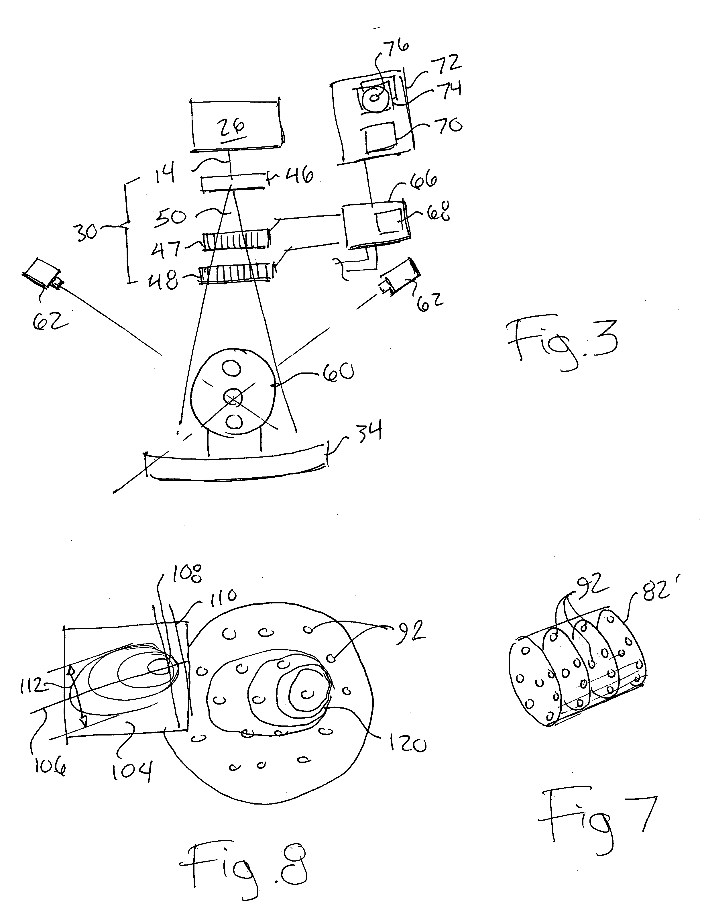
The present invention provides a phantom that can characterize the energy and range of protons using a tissue-mimicking material and an array of intensity detecting elements. The phantom can be used to calibrate a proton or heavy ion therapy machine and to better characterize a CT image used for treatment planning. The phantom includes a tissue-mimicking support and a set of ion detectors spatially separated within the support to detect the passage of heavy ions through the support. The data processing system receiving signals from the ion detectors deducts the location within the phantom of a Bragg peak of heavy ions passing through the support. The phantom can approximate the actual treatment volume and can be used to calibrate planning CT images. The invention provides for multidimensional characterization of the Bragg peak including range and beam widening.

Problems solved by technology

Unfortunately, unlike x-rays, heavy ions are not easily characterized by entrance dose monitors and portal imaging device.
Portal imaging systems, which rely on radiation exiting the patient, are of limited value in a proton imaging system in which the protons stop within the patient.

Method used


Image

  • Phantom for ion range detection
  • Phantom for ion range detection
  • Phantom for ion range detection

Examples


Embodiment Construction

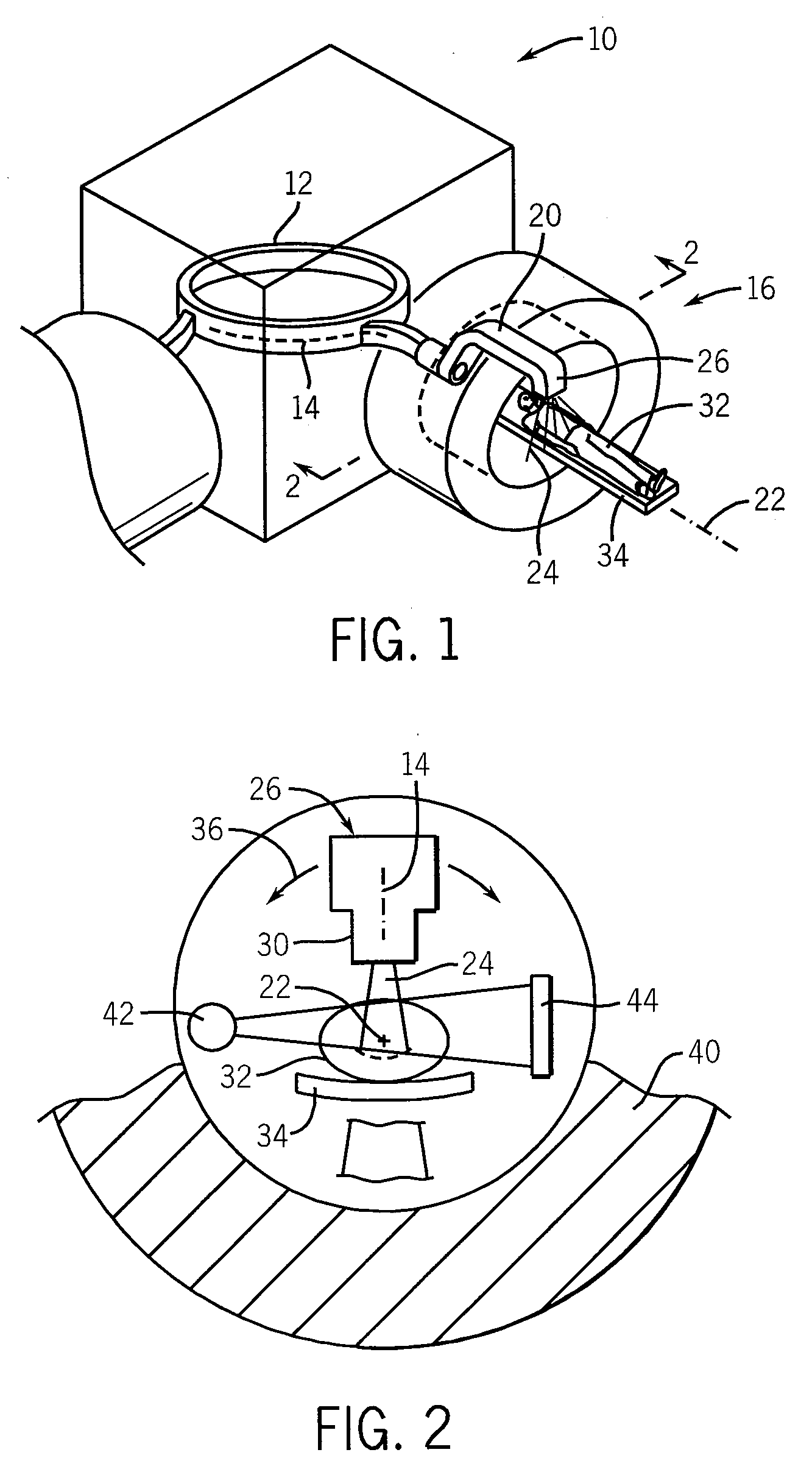
[0026]Referring now to FIG. 1, a heavy ion therapy system 10 may include a cyclotron or synchrotron 12 or other proton source providing a pencil beam of protons 14 to a gantry unit 16.

[0027]Referring also to FIG. 2, the proton beam 14 may be received along an axis 22 into an axial portion of a rotating arm 20 rotating about the axis 22. The rotating arm 20 includes guiding magnet assemblies of a type known in the art to bend the proton beam 14 radially away from the axis 22 then parallel but spaced from the axis 22 to a treatment head 26. The treatment head 26 orbits about the axis 22c with rotation of the rotating arm 20 bending the proton beam 14 back toward the axis 22.

[0028]The treatment head 26 may include a modulation assembly 30 for forming the proton beam 14 into a wider treatment beam (for example a fan or cone beam) and for modulating rays of the beam in energy and intensity to produce a modulated treatment beam 24 as will be described.

[0029]Referring still to FIG. 2, a pa...


PUM

PropertyMeasurementUnit
CTaaaaaaaaaa
energyaaaaaaaaaa
computed tomographyaaaaaaaaaa

Abstract

A phantom for heavy ion radiation therapy provides characterization of an ion beam that may enter but not exit from the phantom. The phantom may include multiple materials and multiple spatially dispersed ion detectors to obtain signals that may be fit to known beam curves to accurately characterize the location and other parameters of Bragg peak of a given ion beam within a patient.

Description

CROSS REFERENCE TO RELATED APPLICATION[0001]This application claims the benefit of U.S. Provisional Application 60 / 891,859, filed Feb. 27, 2007, the disclosure of which is incorporated herein by reference.BACKGROUND OF THE INVENTION[0002]The present invention relates to radiotherapy systems using heavy ions for the treatment of cancer and the like and, in particular, to a phantom for such systems.[0003]External beam radiation therapy may treat a tumor within the patient by directing high-energy radiation in one or more beams toward the tumor. Recent advance external beam radiation systems, for example, as manufactured by Tomotherapy, Inc., treat the tumor with multiple x-ray fan beams directed at the patient over an angular range of 360°. Each of the beams is comprised of individually modulated rays whose intensities can be controlled so that the combined affect of the rays over the range of angles provides an arbitrarily complex treatment area with minimized skin dose.[0004]The ben...

Claims


Application Information

Patent Timeline
Patent Type & Authority Applications(United States)
IPC IPC(8): A61N5/00G01N23/00
CPCA61B6/583A61N2005/1087A61N2005/1076A61N5/1048A61B6/032A61N5/1075
Inventor MACKIE, THOMAS R.FLYNN, RYAN T.
Owner WISCONSIN ALUMNI RES FOUND
Learn More