
Volumetric image formation from optical scans of biological tissue with multiple applications including deep brain oxygenation level monitoring

Inactive Publication Date: 2008-07-24
O2 MEDTECH
View PDF21 Cites 68 Cited by
  • Summary
  • Abstract
  • Description
  • Claims
  • Application Information

AI Technical Summary

Benefits of technology

[0004]However, the types of infrared measurements that are sufficiently developed for practical and everyday medical use are basically directed to “aggregate” or “average” measurement of a biological property over relatively large areas of tissue. It would be desirable to provide for non-invasive infrared measurement of biological tissue sufficiently localized such that actual spatial maps of the biological property or condition are provided that can be expressed as viewable medical images, or medical images suitable for further processing or other uses, while maintaining the safety, low cost, and relative simplicity associated with current “aggregate” or “average” infrared measurement devices.
[0006]Hospital emergency rooms represent another real-world need for fast, safe volumetric imaging of chromophore concentrations, in particular oxygenated hemoglobin levels for the deep human brain. It is often the case, for example, that a patient will arrive in the emergency room in a disoriented and confused state. This condition can have a variety of causes ranging from relatively benign causes, such as drug side effects, to critical and time-sensitive causes, such as stroke or hematoma. It would be desirable to quickly, safely, and inexpensively provide a three-dimensional blood oxygenation map of the patient's brain so that the emergency room physician can (a) take prompt action if the deep brain blood oxygenation map is indicative of a stroke, hematoma, etc., while at the same time (b) prevent excessive equipment and personnel costs if the blood oxygenation map is normal and therefore indicative of a more benign cause for the patient's condition.

Problems solved by technology

Additional issues that may arise in patient monitoring contexts include integrating the hardware and / or software associated with non-invasive three-dimensional mapping of oxygenation levels with more conventional non-invasive monitoring functionalities such as pulse oximetry functionalities.

Method used


Image

  • Volumetric image formation from optical scans of biological tissue with multiple applications including deep brain oxygenation level monitoring
  • Volumetric image formation from optical scans of biological tissue with multiple applications including deep brain oxygenation level monitoring
  • Volumetric image formation from optical scans of biological tissue with multiple applications including deep brain oxygenation level monitoring

Examples


Embodiment Construction

[0025]Hemoglobin, the molecule that carries oxygen in the blood, can exist an oxygenated state, which is designated herein as HbO, and a deoxygenated state, designated herein as Hb. Total hemoglobin, which can be designated as “total Hb” or “HbT”, refers to the collection of the oxygenated and deoxygenated states of hemoglobin (total Hb=HbT=HbO+Hb). Total hemoglobin concentration, which is designated herein by the symbol [HbT], refers to the amount of hemoglobin per unit of blood, and is often expressed in grams per deciliter (g / dl). Similarly, oxygenated hemoglobin concentration, which is designated herein by the symbol [HbO], refers to the amount of oxygenated hemoglobin per unit of blood, and deoxygenated hemoglobin concentration, which is designated herein by the symbol [Hb], refers to the amount of deoxygenated hemoglobin per unit of blood, and both quantities can likewise be expressed in grams per deciliter (g / dl).

[0026]Oxygen saturation refers to the fraction (which can be st...


PUM


Abstract

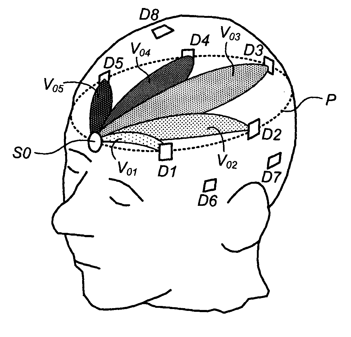
Methods, systems, and related computer program products for non-invasive monitoring of a biological volume, such as a human brain, are described. In one preferred embodiment, each of a plurality of optical sources emits optical radiation into the biological volume each of a plurality of optical detectors detects optical radiation impinging thereupon from the biological volume. The optical measurements are processed to compute a requisite property value associated with each source-detector pair. For each source-detector pair, a volumetric basis region corresponding thereto is weighted by the requisite property value, the volumetric basis region being predetermined and representative of an estimated subvolume of the biological volume encountered by optical radiation emitted from that source and propagating to that detector. The weighted volumetric basis regions are accumulated into a volumetric cumulative array, and a display output is generated based at least in part on the volumetric cumulative array.

Description

CROSS-REFERENCE TO RELATED APPLICATIONS[0001]This application claims the benefit of Provisional Application No. 60 / 885,877, filed Jan. 19, 2007, which is incorporated by reference herein. The subject matter of this application is related to the subject matter of US 2006 / 0015021A1, published on Jan. 19, 2006 (hereinafter “Cheng”), which is incorporated by reference herein.FIELD[0002]This patent specification relates to patient monitoring, including the volumetric imaging of chromophore concentrations or other properties of biological tissue using information acquired from non-invasive optical scans thereof, such as near infrared optical scans. Although applicable in a wide variety of contexts, one particularly advantageous use is for deep brain oxygenation level monitoring.BACKGROUND AND SUMMARY[0003]The use of near-infrared light as a basis for the measurement of biological properties or conditions in living tissue is particularly appealing because of its relative safety as compared...

Claims


Application Information

Patent Timeline
IPC IPC(8): A61B5/1455
CPCA61B5/0073A61B5/14553A61B2562/046A61B2562/0233A61B5/6814A61B5/4064
Inventor WANG, SHIH-PINGCHENG, XUEFENGYU, ZENGPIN
Owner O2 MEDTECH
Learn More