
Leadless Implantable Intravascular Electrophysiologic Device for Neurologic/Cardiovascular Sensing and Stimulation

a neurologic/cardiovascular and electrophysiologic technology, applied in the direction of heart stimulators, internal electrodes, therapy, etc., can solve the problems of insufficient blood pumping to sustain life, hampered technology, and prone to cardiac tamponade acute risk, so as to enhance specificity/integrity, reduce noise/artifact, and control blood flow

Active Publication Date: 2008-05-22
UNIVERSITY OF ROCHESTER
View PDF63 Cites 251 Cited by
  • Summary
  • Abstract
  • Description
  • Claims
  • Application Information

AI Technical Summary

Benefits of technology

[0010]The device can react to physiologic signals it senses and provide electrical stimulation in response to the detected physiologic signals. The electrical stimulation can be used for various purposes, such as to regulate the heart and nervous system, including to stimulate the heart to beat or return to a normal rhythm, stimulate the brain back to normal activity during a seizure episode (epilepsy), provide relief of pain by local nerve stimulation, and control blood flow at various parts of the body by local and central nervous system stimulation. The device communicates the information to other implanted and non-implanted devices that can then act on the information received. For example, information can be transmitted to an infusion pump (implanted or external), which uses the information to inject insulin to control diabetes, anti-arrhythmic drugs for heart rhythm control, or release anti-seizure medication when a seizure is detected. The combination of local data transmission via biologic tissue coupled with wireless communication enhances the specificity / integrity (reduction of noise / artifact) of the data being transmitted.
[0011]The device can also be used in the extravascular or perivascular space. In this form, it has an open / flexible ring that can be adjusted, or self-adjusts to provide no pressure or required contact around the vessel or target region. In this form, the device is positioned using minimally invasive instruments that allow for a small surgical incision, through which tools are designed to position the device at the target site. Fiber optic visualization is used to find the target site and placed the device at that position. The extravascular form of the invention does not necessarily have the size limitations required of an intravascular device, whose size is determined by the vessel's diameter. Accordingly, the extravascular embodiment of the device allows for the attachment of probe modules consisting of flexible inert materials that are used for complex sensing, conducting, or form an antennae for larger geographic operations to provide additional functionality. Those attachments may be made by use of a string or coil and can be placed for easy removal without requiring removal of the perivascular device.
[0012]The device, when placed within a vessel, can also record the pressure imposed by the vessel wall (pressure transducer) and respond accordingly. For example, specific vessels may constrict in response to physiologic stimuli including blood flow changes within the region. This pressure can be detected by the device and information relayed to a controller or another similar device. In addition, the devices can be networked with one another and with an optional controller, and their stimulation pulses or energy delivery coordinated between them to provide the desired response by the biologic tissue. For example, if a seizure is starting in one particular site in the brain, the devices can detect the region and begin to treat while other devices can emit low or high energy signals that are synchronized and protect the normally functioning parts of the brain by making them refractory to the epilepsy electrical activity.

Problems solved by technology

Fibrillation is a condition where the heart has very rapid shallow contractions and, in the case of ventricular fibrillation, may not pump a sufficient amount of blood to sustain life.
However, those technologies are hampered by the use of a transvenous lead for electrophysiologic stimulation.
Those technologies are susceptible to an acute risk of cardiac tamponade, perforation of the heart or vasculature and long term risk of endocarditis or a need for intracardiac extraction of the lead due to failure.
Also, current technologies present a problem for intracardiac defibrillation implantation in younger patients or in patients who are not candidates for the implantation because of anatomical abnormalities.
Complex steps and risks are involved in obtaining venous vascular access and placement of the transvenous lead in the patient population requiring the defibrillation.
Delivering electrical sensing / simulation wires (leads) to specific areas of the brain is difficult.
The risk of infection is high because of the resultant contact of the inner brain areas with areas close to the surface of the skin via the lead.
In addition, normal brain tissue is disrupted in the process, and any complication or infection requires that the whole system be removed.

Method used


Image

  • Leadless Implantable Intravascular Electrophysiologic Device for Neurologic/Cardiovascular Sensing and Stimulation
  • Leadless Implantable Intravascular Electrophysiologic Device for Neurologic/Cardiovascular Sensing and Stimulation
  • Leadless Implantable Intravascular Electrophysiologic Device for Neurologic/Cardiovascular Sensing and Stimulation

Examples


Embodiment Construction

[0023]In describing a preferred embodiment of the invention illustrated in the drawings, certain specific terminology will be used for the sake of clarity. However, the invention is not intended to be limited to that specific terminology, and it is to be understood that the terminology includes all technical equivalents that operate in a similar manner to accomplish the same or similar result.

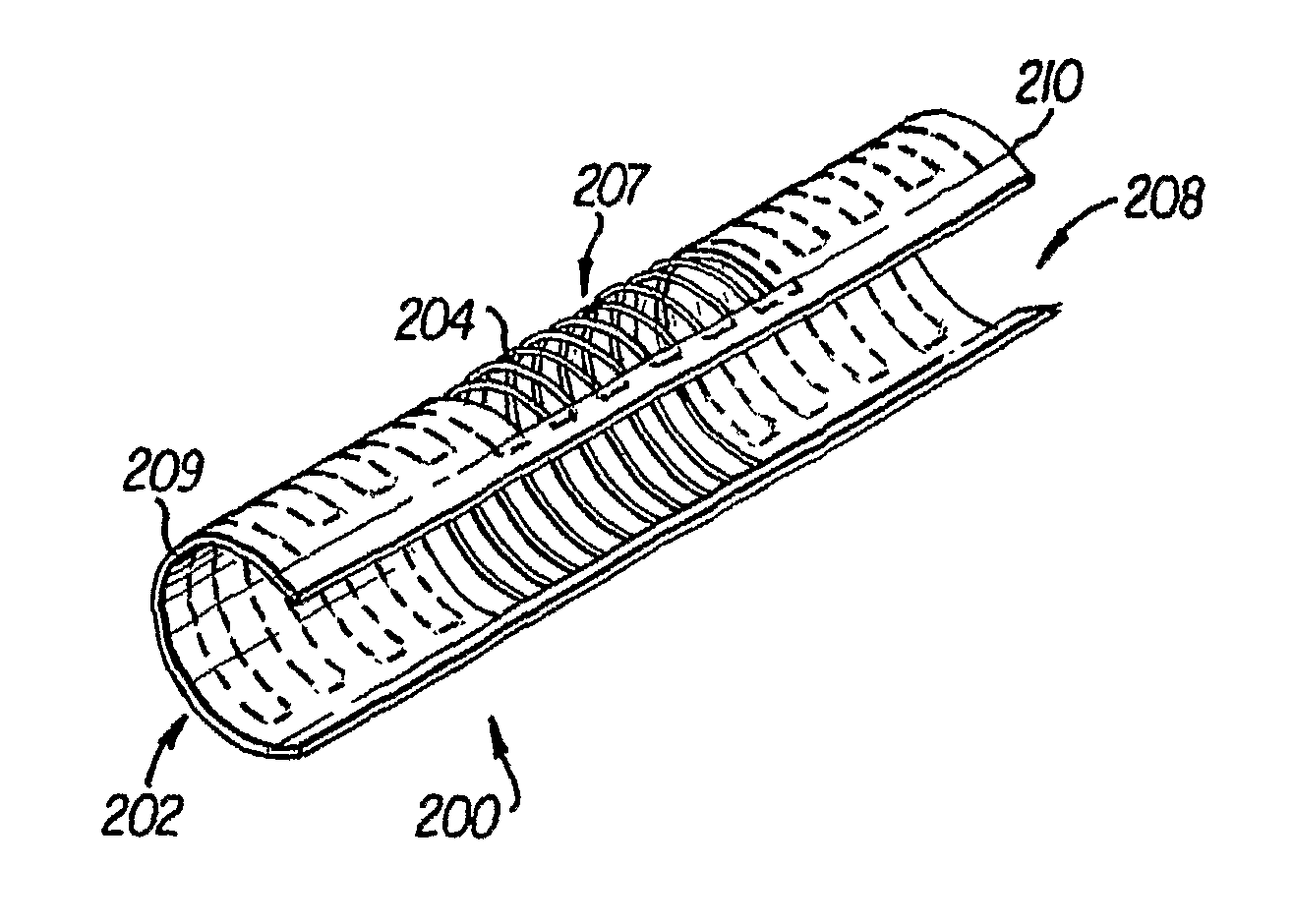
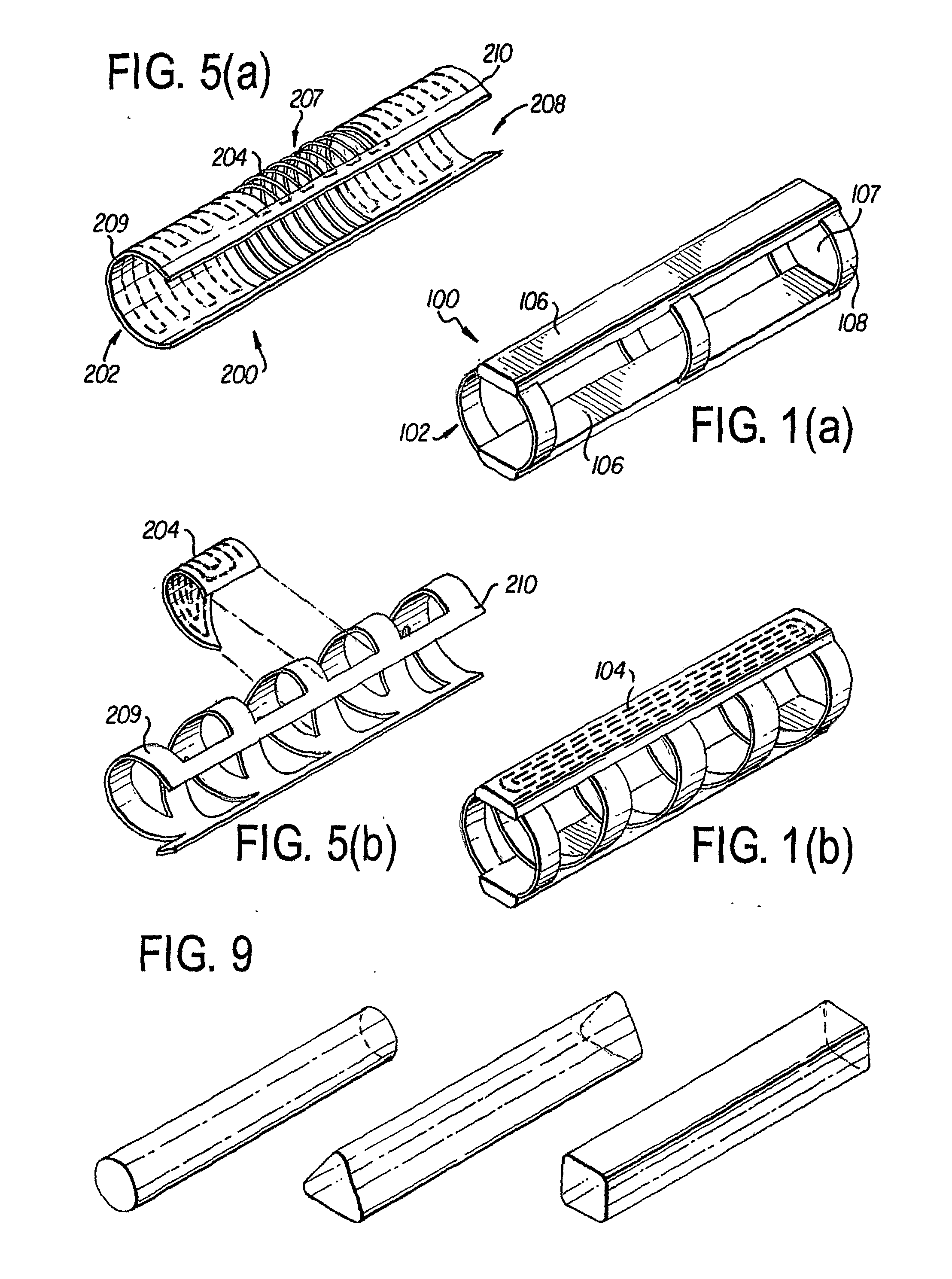
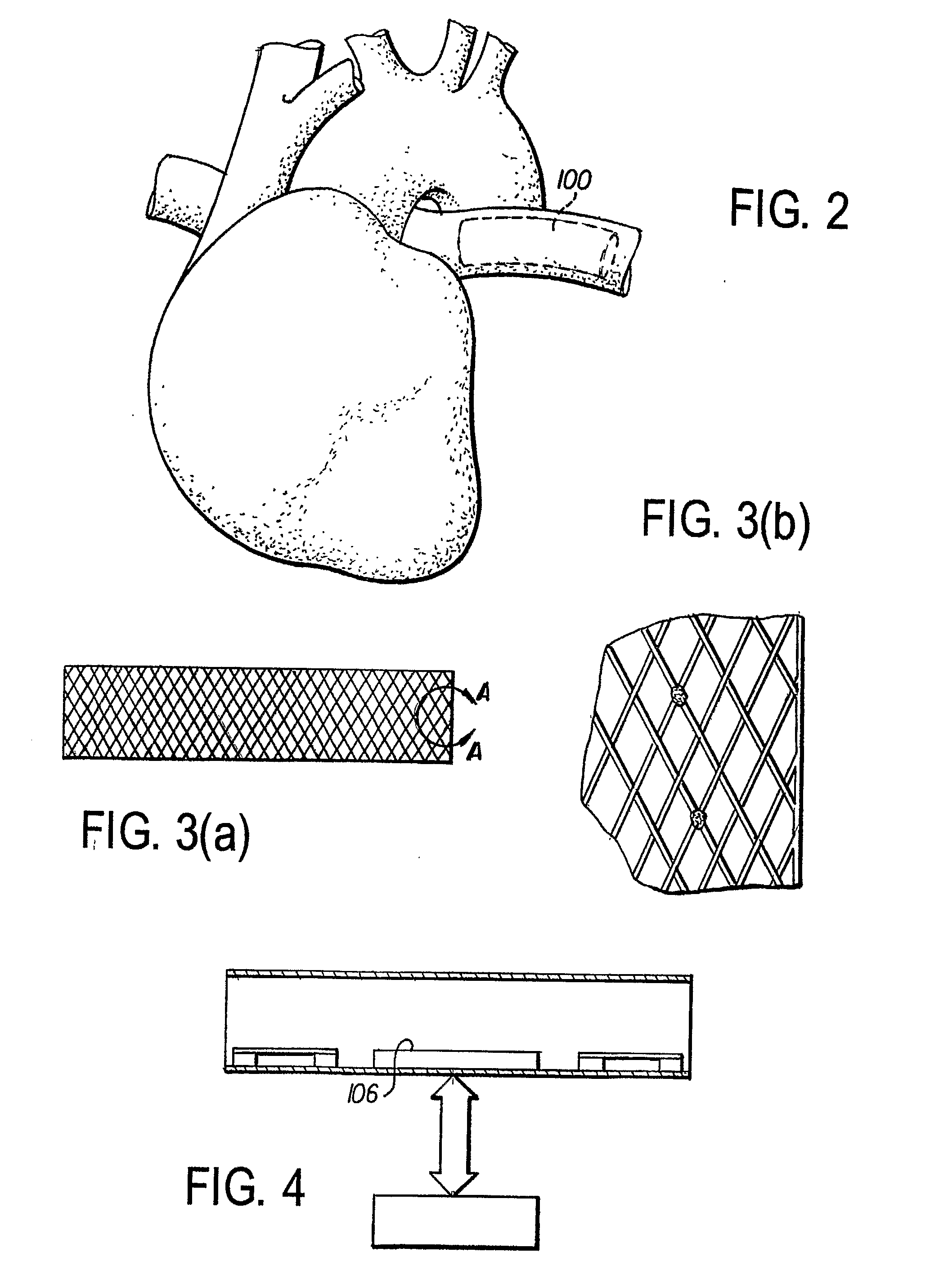
[0024]Referring to the drawings, FIGS. 1(a), (b) and 5(a), (b) show sensing and stimulation devices 100, 200 in accordance with preferred embodiments of the invention. The devices 100, 200 are generally tubular in shape and has a support structure 102, 202, an induction power coil 104, 204 built into the support structure 102, 202, and electronic components 106. The device 100 can be intravascular, as shown in FIGS. 1-4, or extravascular, as shown in FIGS. 5-8. Though the device 100, 200 is shown as having an elongated round tubular shape, it can also have a triangular or square shape, as shown...


PUM


Abstract

A leadless intravascular sensor (100, 200) uses the body tissue as a communication medium. The implantable intravascular device has a tubular stent-like structure (102) for intravascular fixation with embedded microcircuits to allow bipolar and unipolar sensing of cardiac and neurologic electrical activity, sensing of other physiologic signals, local electrical stimulation (cardiac pacing and defibrillation; neurologic stimulation and seizure therapy) as well as the ability to communicate with other implanted and non implanted devices via radio frequency and / or optical communication and / or analog signal communication using the body tissue as the conducting medium. The device can also be used in the extravascular or perivascular space. In this form, it has an open / flexible ring that can be adjusted, or self-adjusts to provide no pressure or required contact around the vessel or target region.

Description

RELATED APPLICATIONS[0001]The present application claims priority to provisional application Ser. Nos. 60 / 567,447, 60 / 567,448 and 60 / 567,449, each of which were filed on May 4, 2004.TECHNICAL FIELD[0002]The present invention is generally related to intravascular and extravascular devices for sensing and stimulation. More particularly, to an implantable intravascular device that allows sensing of cardiac, neurologic and physiologic activity, provides local electrical stimulation, and communicates with other implantable and non-implantable devices via leadless communication using radiofrequency communication, ultrasonic communication, and / or the body tissue as a conducting medium.BACKGROUND OF THE INVENTION[0003]The use of implantable medical devices to treat a variety if diseases is growing due to the rapid advances in technology. Diseases that disrupt the ability of the heart, brain, or nervous system to communicate or function normally include heart rhythm disorders such as ventric...

Claims


Application Information

Patent Timeline
IPC IPC(8): A61N1/05A61N1/08A61N1/372A61N1/375A61N1/39
CPCA61N1/05A61N1/362A61N1/372A61N1/3756A61N1/37288A61N1/375A61N1/37205A61N1/37516
Inventor ROSERO, SPENCER
Owner UNIVERSITY OF ROCHESTER
Learn More