System and method for computer aided detection and diagnosis from multiple energy images

a technology of multiple energy images and computer aided detection, applied in the field of x-ray systems and methods, can solve the problems of difficult visual assessment, calcification, subtle rib fractures, etc., and achieve the effect of improving the accuracy of visual assessment and detecting the condition on a standard chest x-ray

Inactive Publication Date: 2008-02-07
GENERAL ELECTRIC CO
View PDF51 Cites 38 Cited by
  • Summary
  • Abstract
  • Description
  • Claims
  • Application Information

AI Technical Summary

Benefits of technology

[0003] This invention generally relates to computer aided detection and diagnosis (CAD) of an image set. More particularly, this invention relates to a method and system for computer aided detection and diagnosis of dual energy (“DE”) or multiple energy images, as well as of radiographic images, computed tomography images, and tomosynthesis images.

Problems solved by technology

In addition, due to the underlying and overlying structures relevant to the pathologies of interest, visual assessment can be difficult.
Subtle rib fractures, calcifications, and metastatic bone lesions (metastases) in the chest can be difficult to detect on a standard chest X-ray.

Method used


Image

  • System and method for computer aided detection and diagnosis from multiple energy images
  • System and method for computer aided detection and diagnosis from multiple energy images
  • System and method for computer aided detection and diagnosis from multiple energy images

Examples


Embodiment Construction

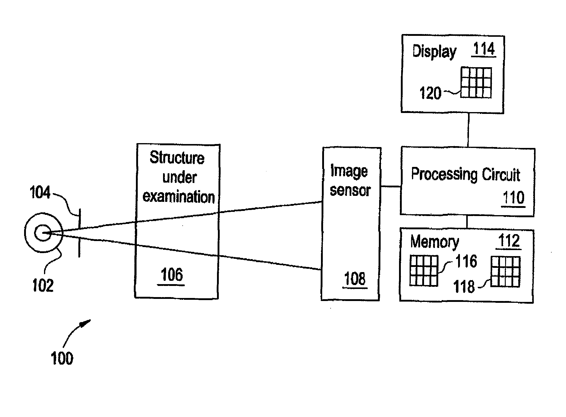
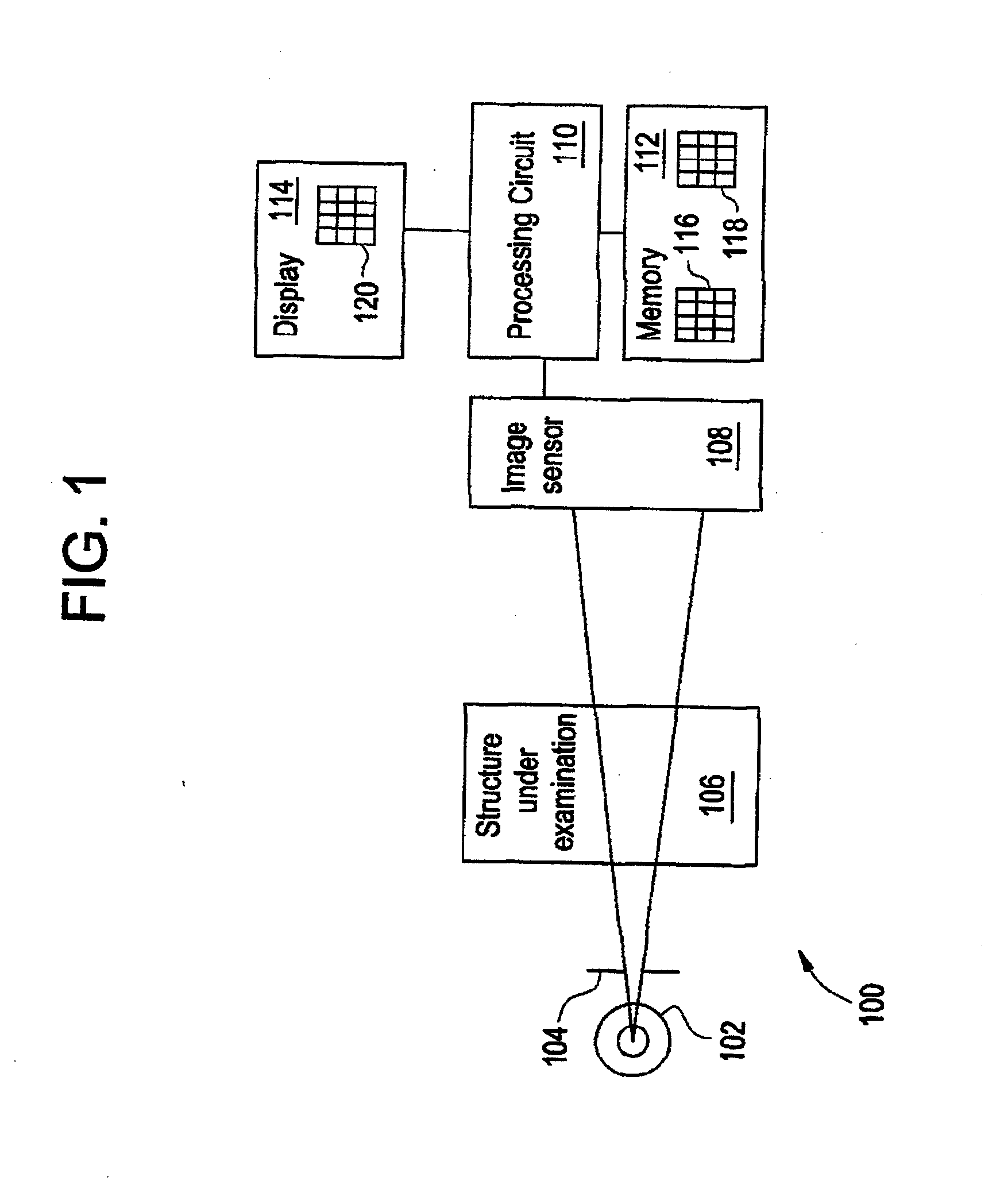
[0032]FIG. 1 illustrates an exemplary X-ray imaging system 100. The imaging system 100 includes an X-ray source 102 and a collimator 104, which subject structure under examination 106 to X-ray photons. As examples, the X-ray source 102 may be an X-ray tube, and the structure under examination 106 may be a human patient, test phantom or other inanimate object under test. The X-ray source 102 is able to generate photons at a first energy level and at least a second energy level different than the first energy level. Multiple, more than two, energy levels are also within the scope of this method and system.

[0033] The X-ray imaging system 100 also includes an image sensor 108 coupled to a processing circuit 110. The processing circuit 110 (e.g., a microcontroller, microprocessor, custom ASIC, or the like) is coupled to a memory 112 and a display 114. The display 114 may include a display device, such as a touch screen monitor with a touch-screen interface. As is known in the art, the s...


PUM


Abstract

A method, system, and storage medium for computer aided processing of an image set includes employing a data source, the data source including an image set acquired from X-ray projection imaging, x-ray computed tomography, or x-ray tomosynthesis, defining a region of interest within one or more images from the image set, extracting feature measures from the region of interest, and reporting at least one of the feature measures on the region of interest. The method may be employed for identifying bone fractures, disease, obstruction, or any other medical condition.

Description

CROSS-REFERENCE TO RELATED APPLICATIONS [0001] This application is a continuation of and claims priority to U.S. patent application Ser. No. 10 / 065,854, filed on Nov. 26, 2002, the disclosure of which is incorporated herein by reference.BACKGROUND OF THE INVENTION [0002] This disclosure relates generally to X-ray systems and methods, and more particularly to a system and method of determining the exposed field of view in an X-ray radiograph. [0003] This invention generally relates to computer aided detection and diagnosis (CAD) of an image set. More particularly, this invention relates to a method and system for computer aided detection and diagnosis of dual energy (“DE”) or multiple energy images, as well as of radiographic images, computed tomography images, and tomosynthesis images. [0004] The classic radiograph or “X-ray” image is obtained by situating the object to be imaged between an X-ray emitter and an X-ray detector made of photographic film. Emitted X-rays pass through th...

Claims


Application Information

Patent Timeline
Patent Type & Authority Applications(United States)
IPC IPC(8): G06K9/00A61B6/00A61B5/00A61B6/03G06F7/40G06F17/50G06K9/34G06T1/00G06T3/00G06T5/30G06T7/00G06T11/00G16H30/20G16H50/20
CPCA61B6/482G06F19/321G06T2211/408G06T11/005G06T2207/30008G06T7/0012A61B6/032A61B6/502A61B6/505G16H50/20G16H30/20A61B6/03
Inventor UPPALURI, RENUKAAVINASH, GOPAL BILIGERITHOMAS, CARSON HALESABOL, JOHN MICHAELJABRI, KADRI NIZARRADER, AMBER ELAINE
Owner GENERAL ELECTRIC CO
Why Eureka
Try Eureka