
Detection of focal epileptiform activity

a focal epileptiform and activity technology, applied in the field of focal epileptiform activity detection, can solve the problems of inability to verbalize an aura, increase the complexity of the signal during a seizure, and disturbed brain function and the resulting eeg

Inactive Publication Date: 2008-01-24
GENERAL ELECTRIC CO
View PDF15 Cites 19 Cited by
  • Summary
  • Abstract
  • Description
  • Claims
  • Application Information

AI Technical Summary

Benefits of technology

[0023]Having the above-mentioned advantages of EEG-based monitoring methods, the mechanism of the invention provides an efficient and a low-cost screening tool by which patients requiring in-depth studies may be selected for imaging or radiological examinations. A further advantage of the invention is that the detection and localization algorithm does not require high computational power, which makes it suitable for various devices with limited computing power, such as ambulatory devices.
[0024]Another aspect of the invention is that of providing an apparatus for detecting focal epileptiform activity. The apparatus includes a measurement module configured to obtain a first plurality of brain wave signals from a subject and a first computing module configured to determine a signal-specific measure indicative of the degree of epileptiform activity for at least some of the first plurality of brain wave signals, whereby a second plurality of signal-specific measures are obtained. The apparatus further includes an indicator module configured to provide an indication of the presence of focal epileptiform activity based on the second plurality of signal-specific measures.
[0025]In a still further embodiment, the invention provides a computer program comprising a first program code portion configured to determine a signal-specific measure indicative of the degree of epileptiform activity for a plurality of brain wave signals, thereby to obtain a corresponding plurality of signal-specific measures and a second program code portion configured to provide an indication of the presence of focal epileptiform activity based on the second plurality of signal-specific measures. It is thus to be noted that since a conventional measurement device may be upgraded by a plug-in unit that includes software enabling the measurement device to detect focal epileptiform activity, the plug-in unit does not necessarily have to take part in the acquisition of the brain wave signal data.

Problems solved by technology

Different derangements of internal system homeostasis disturb the environment in which the brain operates, and therefore the function of the brain and the resulting EEG are disturbed.
However, when brain activity is recorded from the scalp, the measured signal is a composition originating from multiple sources, and methods indicative of the complexity of the signal show an increase during a seizure, cf.
However, young children, for example, may be unable to verbalize an aura, and the aura or focal onset of a seizure may also be so brief that it may not be noticed before a secondary generalization of the seizure occurs.
However, identification of epileptic seizures of ICU patients is difficult, since the unconsciousness of the patients may be generated either by sedative drugs or by a brain disorder, such as an epileptic seizure.
However, imaging techniques are not 100% sensitive to neither type of lesions.
Furthermore, functional imaging is cumbersome to perform.
In a typical clinical environment, imaging can be performed maximally once per day, because of resource limitations.
However, these monitors provide only binary information about epileptiform activity, i.e. they provide information only about the presence or absence of epileptiform activity.
Due to the binary information utilized, the monitors are not efficient for detecting focal epileptiform activity or for localizing the focus of epileptiform activity.
This drawback is due to the fact that focal epileptiform activity may be observable in large brain areas and may evolve even to the other hemisphere.

Method used


Image

  • Detection of focal epileptiform activity
  • Detection of focal epileptiform activity
  • Detection of focal epileptiform activity

Examples


Embodiment Construction

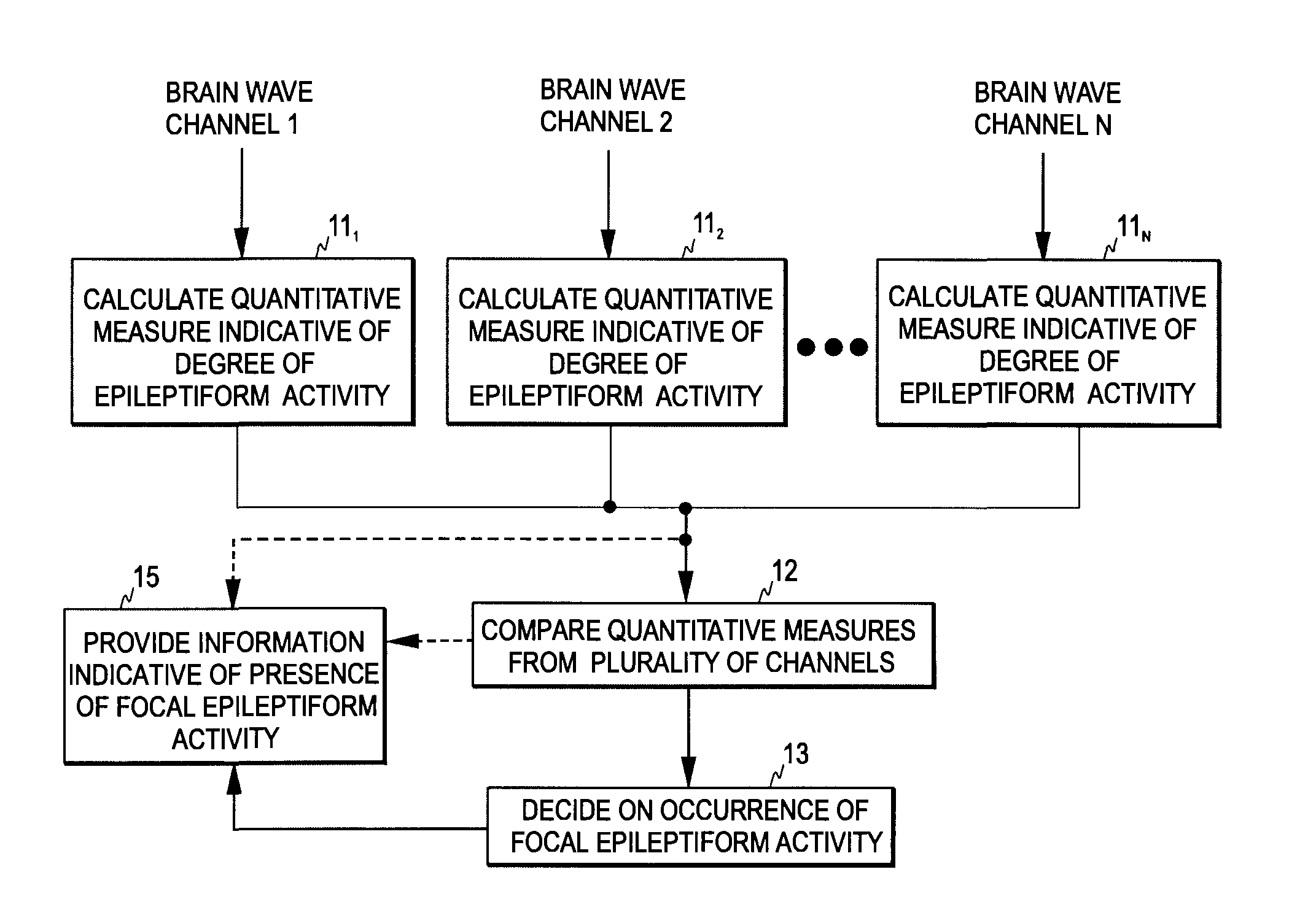
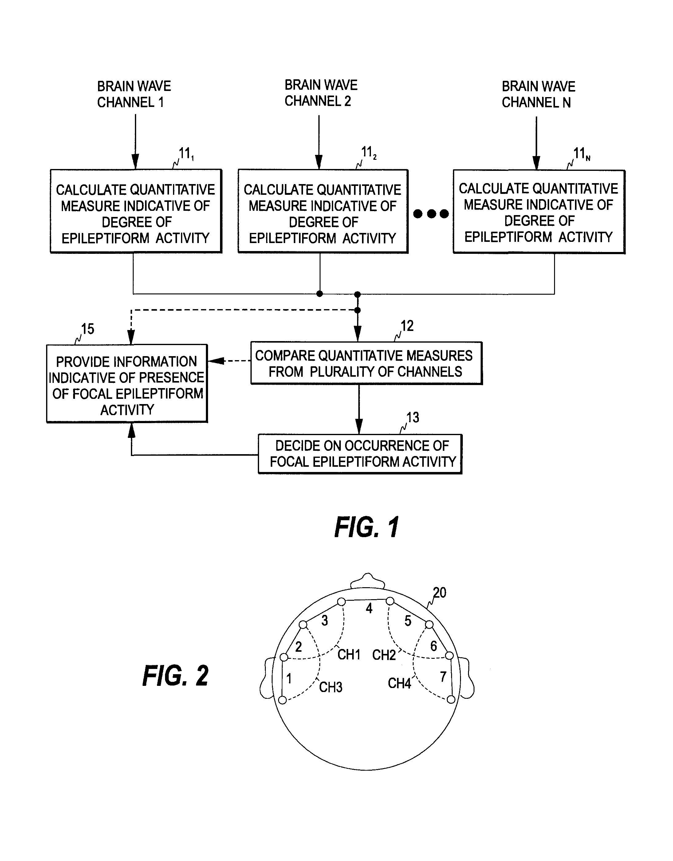
[0036]FIG. 1 is a flow diagram illustrating one embodiment of the method of the invention. In the present invention, N (N=2, 3, . . . ) brain wave signals are measured from a subject. As is common in the art, each incoming brain wave signal is sampled and the digitized signal samples are processed as sets of sequential signal samples representing finite time blocks or time windows, commonly termed “epochs”. Here, each brain wave signal is also referred to as a channel, i.e. each brain wave signal is obtained through a corresponding channel.

[0037]Based on each brain wave signal, a measure indicative of the degree of epileptiform activity is determined (steps 111, . . . , 11N), whereby N signal-specific values are obtained for the measure in each time window. As mentioned above, the measure here refers to a quantitative measure of epileptiform activity, which changes in a monotonic manner on a continuous scale according to the changes in the epileptiform activity.

[0038]Next, some or a...


PUM


Abstract

The invention relates to detection of focal epileptiform activity. In order to accomplish an EEG-based mechanism that enables the detection and localization of focal epileptiform activity, a first plurality of brain wave signals is obtained from a subject and a signal-specific measure indicative of the degree of epileptiform activity is determined for at least some of the first plurality of brain wave signals, thereby to obtain a second plurality of signal-specific measures. An indication of the presence of focal epileptiform activity is provided based on the second plurality of signal-specific measures. This may involve, for example, a comparison of the signal-specific measures. If significant mutual differences are detected in the measures, focal epileptiform activity is detected.

Description

FIELD OF THE INVENTION [0001]The present invention relates generally to the detection of focal epileptiform activity.BACKGROUND OF THE INVENTION [0002]Electroencephalography (EEG) is a well-established method for assessing brain activity. When measurement electrodes are attached on the skin of the skull surface, the weak biopotential signals generated in brain cortex may be recorded and analyzed. The EEG has been in wide use for decades in basic research of the neural systems of the brain as well as in the clinical diagnosis of various central nervous system diseases and disorders.[0003]The EEG signal represents the sum of excitatory and inhibitory potentials of large numbers of cortical pyramidal neurons, which are organized in columns. Each EEG electrode senses the average activity of several thousands of cortical pyramidal neurons.[0004]The EEG signal is often divided into four different frequency bands: Delta (0.5-3.5 Hz), Theta (3.5-7.0 Hz), Alpha (7.0-13.0 Hz), and Beta (13.0-...

Claims


Application Information

Patent Timeline
IPC IPC(8): A61B5/04
CPCA61B5/04017A61B5/726A61B5/4094A61B5/0476A61B5/316A61B5/369A61B5/374
Inventor SARKELA, MIKA
Owner GENERAL ELECTRIC CO
Learn More