
Method and apparatus for the determination of intrinsic spectroscopic tumor markers by broadband-frequency domain technology

Inactive Publication Date: 2008-01-10
RGT UNIV OF CALIFORNIA
View PDF5 Cites 49 Cited by
  • Summary
  • Abstract
  • Description
  • Claims
  • Application Information

AI Technical Summary

Benefits of technology

[0019] The proposed optical method can be applied to women of all ages, is not painful and the result of the analysis can be interpreted by a computer algorithm. The instrumentation does not produce harmful radiation and it can be installed in a doctor's cabinet. The instrumentation is on order of magnitude less expensive that the conventional mammography method.

Problems solved by technology

Despite years of research, the promise of non-invasive optical biopsy of breast tumors has not been fully realized.
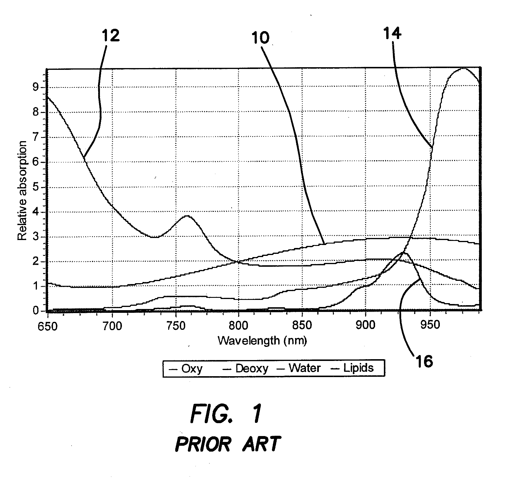
The challenge of spectroscopy in tissues has been the separation of attenuation due to scattering from that due to tissue absorption.
The problem with determining the absorption in tissues is that the apparent absorption depends on the amount of scattering in the tissue.
None of these approaches can give the detailed wavelength information that is necessary for the applications of the methods described in this disclosure.
None of the work done neither at UCI nor in other laboratories was done with the purpose of identifying specific tumor spectral components.
However, the knowledge of the amount of tissue components is not enough to uniquely identify tumors, even less to distinguish between malignant and benign tumors.
The disadvantage of the available method is that the sensitivity is not 100%.
However, x-ray mammography cannot be applied in young women due to the dense spectroscopic breast from the x-ray point of view.
Furthermore, mammography has limited sensitivity and specificity.
Mammography is also painful and the results must be read by an experienced radiologist.
No automatic method for the identification of tumors from the mammography slide is available.

Method used


Image

  • Method and apparatus for the determination of intrinsic spectroscopic tumor markers by broadband-frequency domain technology
  • Method and apparatus for the determination of intrinsic spectroscopic tumor markers by broadband-frequency domain technology
  • Method and apparatus for the determination of intrinsic spectroscopic tumor markers by broadband-frequency domain technology

Examples


Embodiment Construction

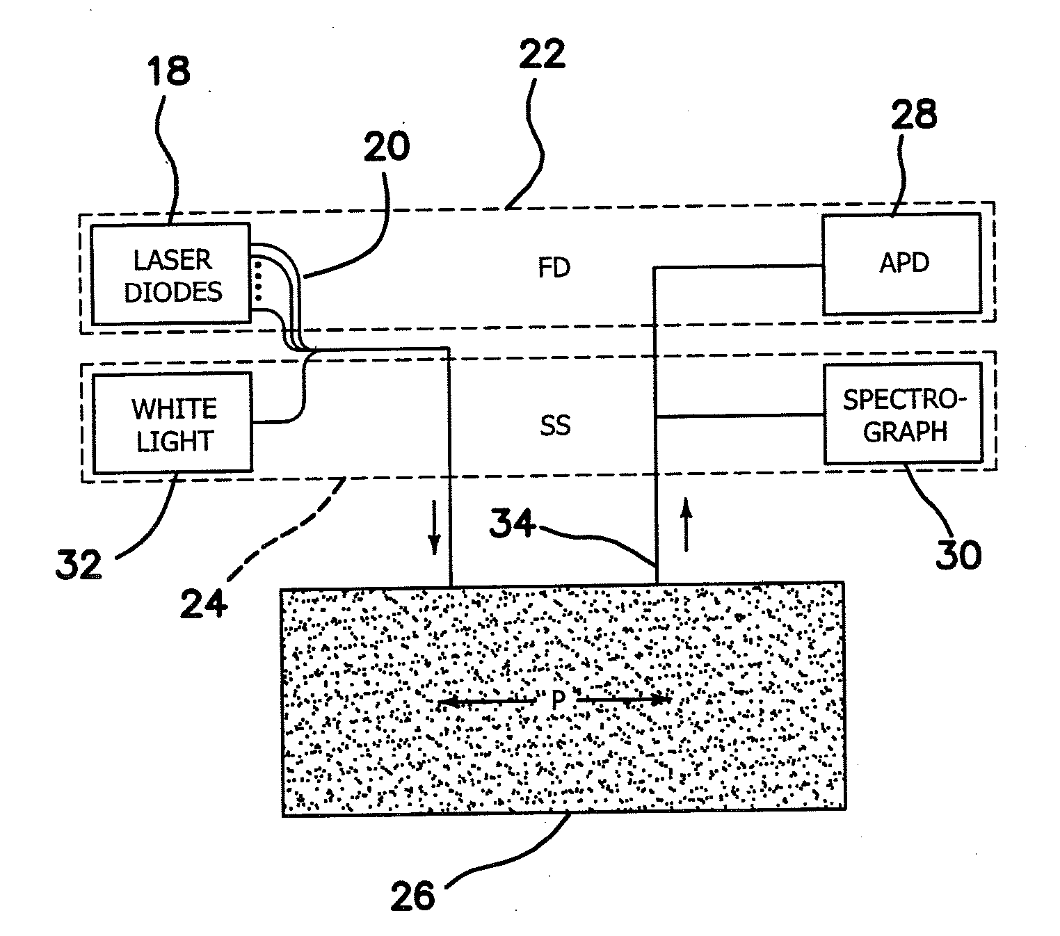
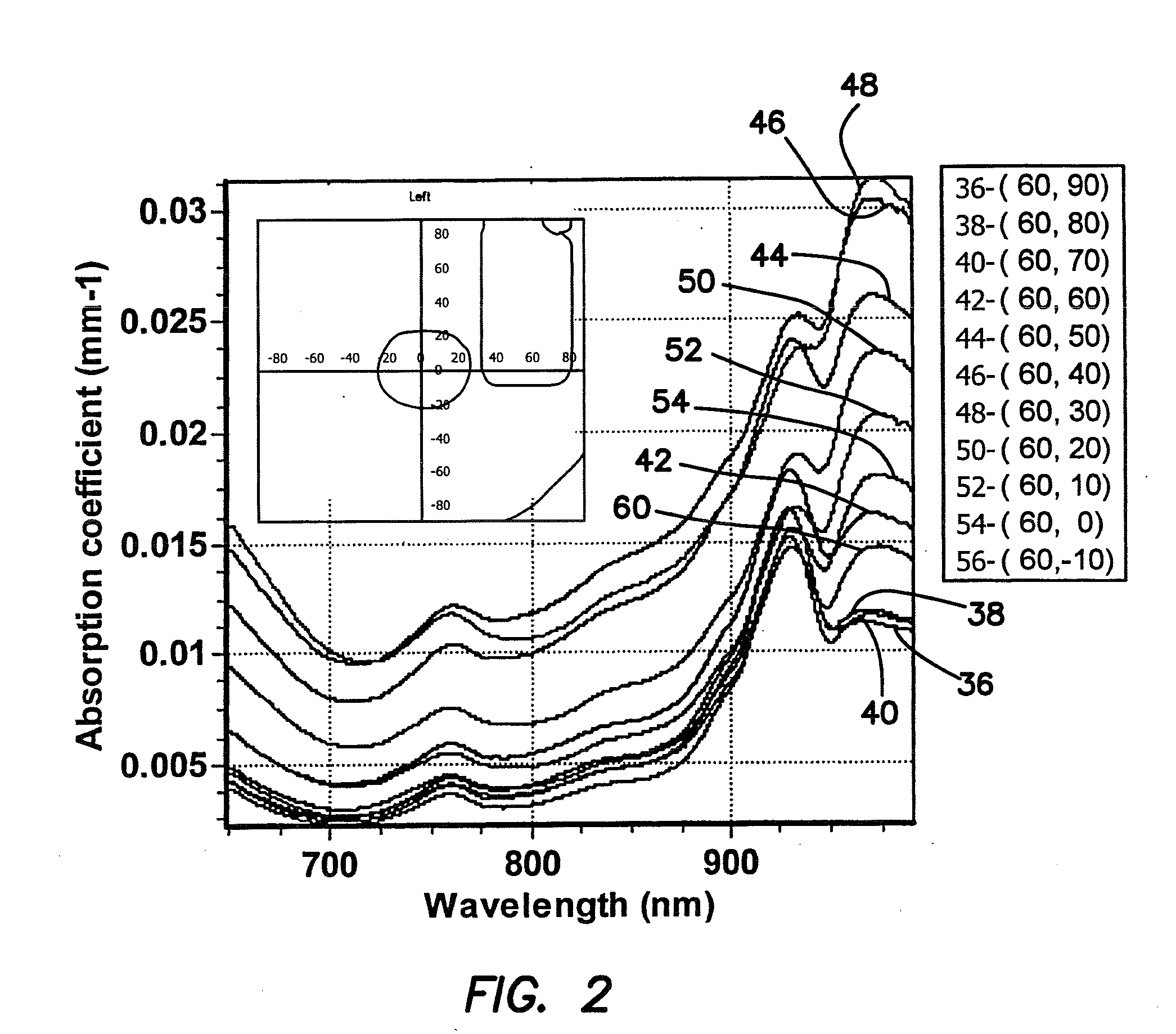
[0071] The illustrated embodiment is a double differential method to analyze the near-infrared spectra of regions of the human breast with tumors. We show that the near-infrared (650-1000 nm) spectra of breasts with tumors have characteristic absorption bands in the lipid fingerprint region that are unaccounted for in conventional spectral models. These spectral components do not appear in the normal breast of the same patient or in regions of the diseased breast away of the tumor. These spectral components originate from lipids that are present in tumors either in different abundance than in the normal breast or new lipid components that are caused by the different metabolism in tumors. Furthermore, the water band in the 980-1000 nm region also shows distinct variations in the tumor region compared to the normal breast. By combining the information in the lipid and water region, we constructed an index that is characteristic of the tumor region (100% specificity and 93% sensitivity...


PUM


Abstract

The illustrated embodiment is an improvement in a method of optically analyzing tissue in vivo in an individual to obtain a unique spectrum for the tissue of the individual, the improvement including the steps of optically measuring the tissue of the individual to obtain a spectrum of an optical parameter, and identifying a spectral signature specific to a metabolic or physiologic state in the tissue of the individual with a unique spectrum for the tissue by considering only the spectral differences between a first metabolic or physiologic state of the tissue of the individual and one or more other metabolic or physiologic states of the tissue of the individual such that identification of the spectral signature is self-referencing with respect to intra-individual metabolic or physiologic variations. The method also includes separating benign and malignant lesions only using the shape or a characteristic of the spectrum.

Description

RELATED APPLICATIONS [0001] The present application is related to U.S. Provisional Patent Application Ser. No. 60 / 747,384, filed on May 16, 2006, which is incorporated herein by reference and to which priority is claimed pursuant to 35 USC 119.GOVERNMENT RIGHTS [0002] This invention was made with Government support under Grant Nos. CA105480 and EB000559, awarded by the National Institutes of Health. The Government has certain rights in this invention.BACKGROUND OF THE INVENTION [0003] 1. Field of the Invention [0004] The invention relates to the field of methods of use of near-IR for the determination of optical parameters of tissues and apparatus for performing the same. [0005] 2. Description of the Prior Art [0006] Despite years of research, the promise of non-invasive optical biopsy of breast tumors has not been fully realized. During the past decade we witnessed a renewed interest in this field due to the realization that quantitative spectroscopy can be performed in thick tissu...

Claims


Application Information

Patent Timeline
IPC IPC(8): A61B6/00
CPCA61B5/0059G01N21/359A61B5/06
Inventor GRATTON, ENRICOKUKRETI, SHWAYTACERUSSI, ALBERT E.TROMBURG, BRUCE J.
Owner RGT UNIV OF CALIFORNIA
Learn More