Radiometric Approach to Temperature Monitoring Using a Magnetic Resonance Scanner

a magnetic resonance scanner and radiometric technology, applied in the direction of measuring using nmr, instruments, applications, etc., can solve the problems of difficult use of such temperature measurement techniques in medical applications, many rf noise sources in medical facilities, and difficult measurement in the radio frequency (rf) region of the electromagnetic spectrum, so as to achieve absolute non-invasive thermal imaging of target tissue volume, improve patient treatment effect, and improve accuracy

Inactive Publication Date: 2007-12-20
THE JOHN HOPKINS UNIV SCHOOL OF MEDICINE
View PDF4 Cites 17 Cited by
  • Summary
  • Abstract
  • Description
  • Claims
  • Application Information

AI Technical Summary

Benefits of technology

[0013] An advantage of the present invention is to provide more accurate, absolute non-invasive thermal imaging of a target tissue volume.
[0014] Another advantage of the present invention is to provide more effective diagnoses of medical ailments such as the identification of tumors.
[0015] Another advantage of the present invention is to provide real time thermal imagery to better assist in the treatment of patients.
[0016] Additional features and advantages of the invention will be set forth in the description which follows, and in part will be apparent from the description, or may be learned by practice of the invention. The objectives and other advantages of the invention will be realized and attained by the structure particularly pointed out in the written description and claims hereof as well as the appended drawings.
[0017] To achieve these and other advantages and in accordance with the purpose of the present invention, as embodied and described, a system for generating thermal imagery using an MR scanner comprises: an RF coil; a tuning means connected to the RF coil; a pre-amp connected to the tuning means; a demodulator connected to the output of the pre-amp; a digitizer connected to the output of the demodulator; and a computer connected to the output of the digitizer, the computer having a computer readable medium encoded with a program for collecting noise signals detected by the RF coil, calculating a variance of the noise signals, and converting the variance to a temperature.
[0018] In another aspect of the present invention, a method for using an MR scanner to measure absolute temperature of a target volume, the method comprises the steps of: tuning an RF coil; collecting a plurality of signal data from the RF coil; determining a variance corresponding to the plurality of signal data; and converting the variance to an absolute temperature data.

Problems solved by technology

However, measurements in the lower frequency (i.e. longer wavelength) regions of the electromagnetic spectrum, such as in the radio frequency (RF) region of the spectrum, can be difficult due to the weakness of the signal relative to system thermal noise and interference from the environment.
However, the problems associated with system noise and RF environmental factors, such as external noise factors, make the use of such temperature measurement techniques difficult in medical applications.
For example, a medical facility presents many RF noise sources that are not encountered by a remote sensing instrument in the depths of space.

Method used


Image

  • Radiometric Approach to Temperature Monitoring Using a Magnetic Resonance Scanner
  • Radiometric Approach to Temperature Monitoring Using a Magnetic Resonance Scanner
  • Radiometric Approach to Temperature Monitoring Using a Magnetic Resonance Scanner

Examples


Embodiment Construction

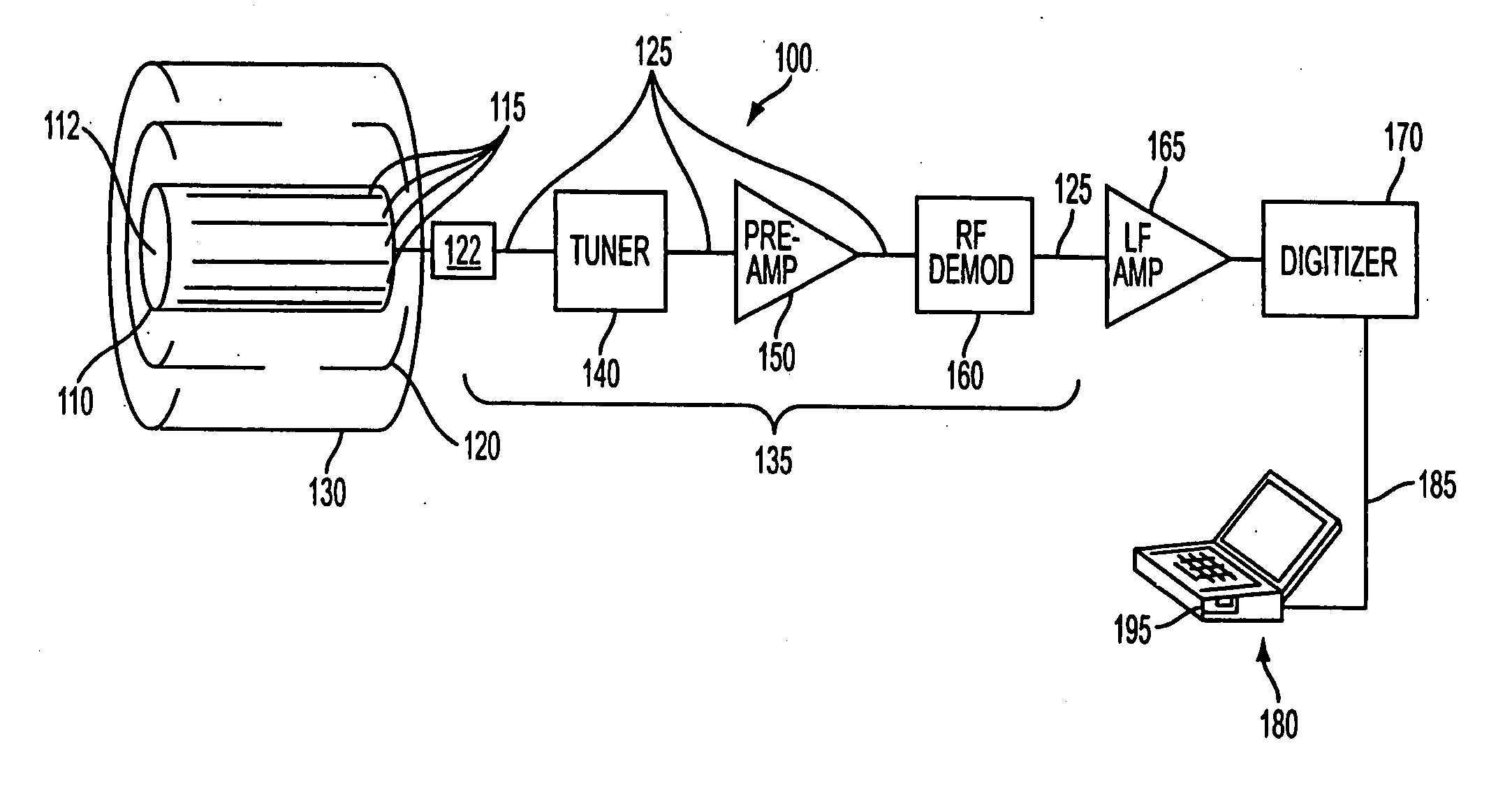
[0032] The present invention uses an MR scanner as a passive RF radiometer to detect thermal noise radiated from a target tissue. It is known that tissue (or any material, for that matter) emits electromagnetic radiation that varies with temperature. In the RF region of the electromagnetic spectrum, the statistical variance of the noise present in the RF radiated power is a function of temperature of the tissue. In an MR scanner, the RF noise that is generated by the tissue in the scanner may induce a current in the RF coil. The induced current in the RF coil may be detected by the electronics of the MR scanner, and processed to produce measurement of the absolute temperature of the tissue. The thermal noise generated by the tissue is a low amplitude signal, and subject to environmental noise and interference. However, by using the MR scanner room for electromagnetic shielding, one may create a sufficiently quiet RF environment around the tissue, enabling accurate measurement of the...


PUM

PropertyMeasurementUnit
frequenciesaaaaaaaaaa
thermalaaaaaaaaaa
temperatureaaaaaaaaaa

Abstract

Disclosed is a method and system for acquiring absolute temperature imagery using an MR scanner. The method involves using the RF coil as a passive antenna, and performing radiometric measurements of the noise variance of the target within the field of view of the RF coil. The noise variance corresponds to the absolute temperature of the volume within the field of view of the RF coil. The room of the MR scanner is used for electromagnetic shielding during the acquisition of radiometric data. This method may be performed with minimal or no add-ons to existing MR scanner hardware. Disclosed are a method for calibrating an MR scanner for radiometric temperature measurements, and a method for acquiring and generating thermal imagery with a calibrated MR scanner.

Description

[0001] This application claims the benefit of U.S. Provisional Patent Application No. 60 / 485,299, filed on Jul. 7, 2003, which is hereby incorporated by reference for all purposes as if fully set forth herein.[0002] Research and development efforts associated with the subject matter of this patent application was supported by the National Institutes of Health under Grant Nos. R01HL61672 and R01HL57483.BACKGROUND OF THE INVENTION [0003] 1. Field of the Invention [0004] The present invention relates to the creation of three-dimensional thermal images using a Magnetic Resonance (MR) imaging system, where the imagery shows absolute temperatures within a target tissue. [0005] Three dimensional imaging of absolute temperature within a target tissue provides great benefit in both medical diagnostics and treatment. In medical diagnostics, it is known that tumors, for example, maintain a different temperature relative to the surrounding tissue. This is particularly true with breast cancer. T...

Claims


Application Information

Patent Timeline
Patent Type & Authority Applications(United States)
IPC IPC(8): A61B5/05G01K11/00G01RG01R33/48
CPCG01K11/006G01R33/4804G01K2213/00G01K13/002G01K13/20
Inventor EL-SHARKAWY, ABDEL-MONEMATALAR, ERGIN
Owner THE JOHN HOPKINS UNIV SCHOOL OF MEDICINE
Why Eureka
Try Eureka