
Filter for separating blood cells

a filter and blood cell technology, applied in the separation process, laboratory glassware, instruments, etc., can solve the problems of difficult to fill glass fiber filters, and glass fiber filters are difficult to fill

Inactive Publication Date: 2007-10-04
FUJIFILM CORP
View PDF14 Cites 25 Cited by
  • Summary
  • Abstract
  • Description
  • Claims
  • Application Information

AI Technical Summary

Benefits of technology

The present invention provides a filter for separating blood cells using a water-insoluble substance fixed to a substrate. The filter has voids that allow blood cells to pass through without clogging, and can recover blood plasma with a high efficiency. The filter can be used in a blood filtering instrument or a blood pre-treatment element. The technical effects of the invention include improved efficiency in separating blood cells and reduced clogging of the filter.

Problems solved by technology

In the blood cell separation apparatus using the above-described glass fibers described above, there was the technical problem that it is difficult to fill sheet-like glass fibers having elasticity in a microstructural portion such as a microchip.
When a glass fiber filter as a filtering material is filled in a microstructural portion of a microchip, it gives priority to fill the same such that a liquid filtered does not leak, and therefore, it is difficult to fill the glass fiber filter such that the voids do not change.
As a result, the glass fiber filter collapses, and voids are liable to become small.
Consequently, where glass fibers as a filtering material are filled in a microstructural portion of a microchip, there was the disadvantage that red blood cells clog in the filtering material, and a sufficient amount of blood plasma cannot be recovered.
Contrary to this, where glass fibers are airily filled such that the glass fibers do not collapse, there was the problem that a space is generated between a cartridge member filling glass fibers, and the glass fibers, and as a result, blood cells pass thought without being captured.

Method used


Image

  • Filter for separating blood cells
  • Filter for separating blood cells
  • Filter for separating blood cells

Examples


example 1

Preparation of Blood Cell Separation Filter Using Resin-Made Micropillars and Result of Blood Filtration

(A) Preparation of Resin-Made Micropillars

[0219] Resin-made micropillars having an equivalent circle diameter of 4 μm or less and being fixed to a substrate with contact points were prepared by the following procedures.

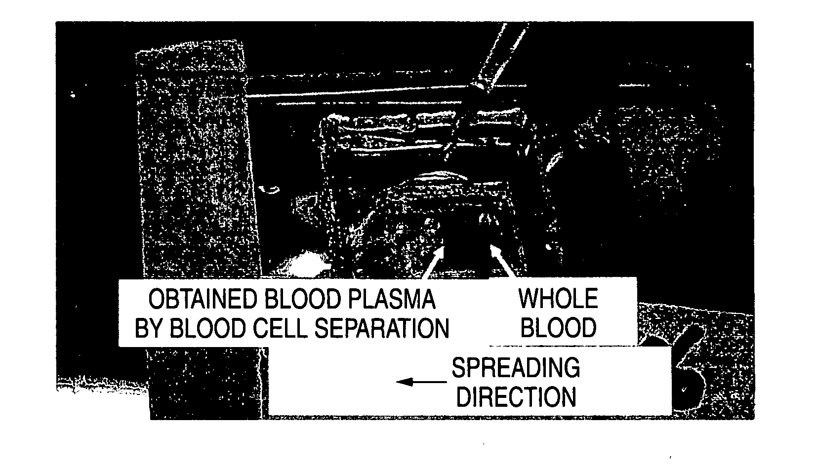
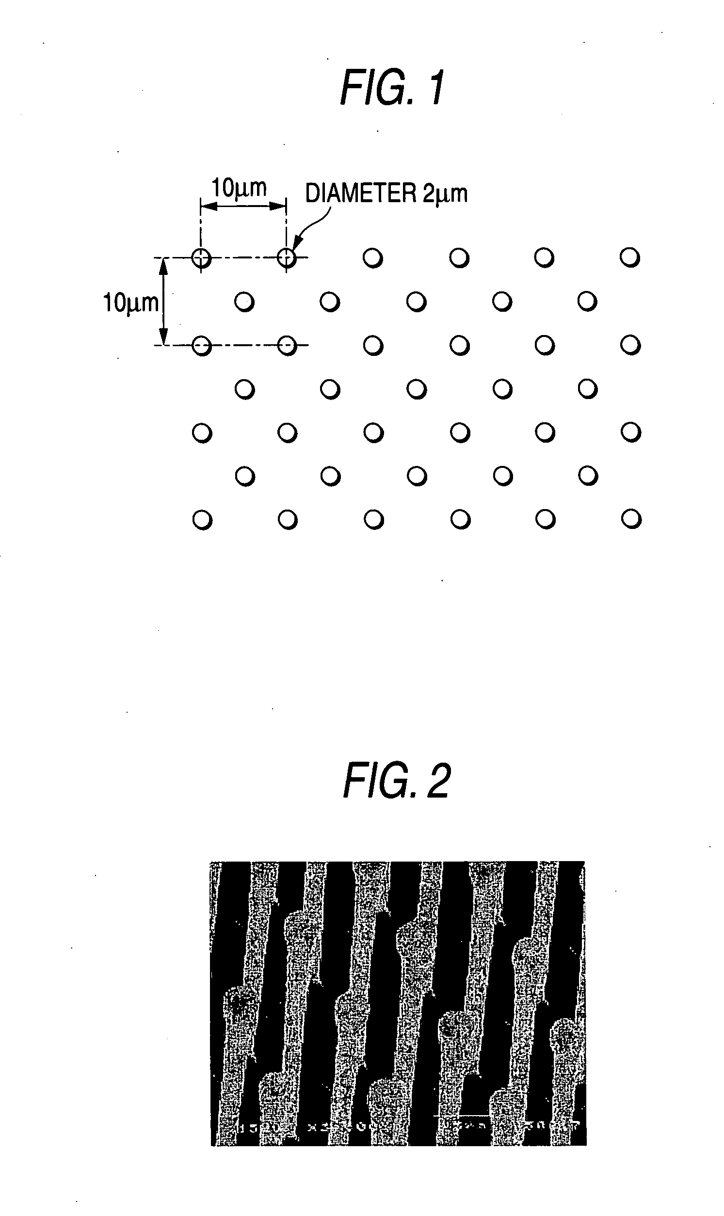
[0220] A silicon wafer was dry etched using a semiconductor processing technology to form wells each having a diameter of 3 μm and a depth of 20 μm at a distance of pitch 10 μm in a checkered pattern over an area having a width of 10 mm and a length of 10 mm. Thus, a mold for use in imprinting was prepared. Using this mold made of a silicon wafer, heat and pressure were applied to a plate of a polystyrene (PS) resin to prepare resin-made micropillars by an imprinting method. The micropillars prepared had a diameter of 2 μm and a height of 27 μm and were arranged in a checkered pattern at a distance of pitch 10 μm. The region on which the pillars could be prepare...

example 2

Capture of Red Blood Cell Using Silicon-Made Micropillars

[0223] Using an apparatus MC-FAN (a product of Hitachi Haramachi Electronics Co., Ltd.) that measures by observing flow of a blood, a shape of a custom chip used for observation of MC-FAN was modified as application of conducting observation of dynamic form when cells such as red blood cells pass through a fine flow passage, thereby preparing a custom chip having provided columnar projections generally called pillars in the flow passage of the chip. Specifically, a silicon wafer was used as a substrate, the silicon wafer was dry etched using a semiconductor processing technology, and the surface of the silicon wafer was further oxidized. Pillars were prepared to have a constant height of 3.5 μm, and a diameter of 3 μm and 4 μm. The pillars were arranged with three lines at a constant pitch of 20 μm toward a direction flowing a blood. Whole blood of a healthy man collected using a heparin lithium blood collecting tube was flow...

example 3

Preparation of Flat Plate Chip (Fitting Type Dry Elemental Device) and its Result

(A) Preparation of Flat Plate Chip (Fitting Type)

[0225] The dry analytical device 50 by fitting the upper member 30 and the lower member 40 shown in FIG. 12 through the porous membrane 52 was prepared according to the following procedures.

[0226] The upper member 30 and the lower member 40, each having a size of about 24 mm×28 mm, molded using a transparent polystyrene were provided. Diameter of the fitting projected portion 35 and the fitting depressed portion 46 was about 9 mm.

[0227] Glass fibers obtained by previously treating a glass filter paper (GF / D, a product of Whatman Co.) for red blood cell capturing and blood plasma extraction and then being subjected to PMEA treatment, as a filtration filter 36 (analytical element) were tightly fixed to the flow passage 34 using a double-sided tape, and filled therein. Length of the glass fibers was apparently longer that an equivalent circular diameter...


PUM

PropertyMeasurementUnit
heightaaaaaaaaaa
distanceaaaaaaaaaa
heightaaaaaaaaaa

Abstract

A filter for separating blood cells, which comprises: a substrate; and at least one water-insoluble substance fixed to the substrate, wherein the at least one water-insoluble substance has an equivalent circle diameter of 4 μm or less and a height equal to or larger than the equivalent circle diameter, wherein blood cells are separated by substantially being captured with the at least one water-insoluble substance; and a blood filtration instrument and a blood analytical device, using the filter for filtration of blood and body fluid.

Description

BACKGROUND OF THE INVENTION [0001] 1. Field of the Invention [0002] The present invention relates to a filter for separating blood cells, used in a blood test method of human being and other animals, and a filtering instrument and an analytical device, using the filter. [0003] 2. Description of the Related Art [0004] A method of diagnosing disease of human being using blood, urine and the like as an analyte has conventionally been conducted for a long time as a method that can diagnose simply without damaging human body. [0005] In particular, the blood enables many examination items to diagnose. [0006] A wet chemistry analytical method has conventionally been developed as an analytical method for examining such many items. This is a method using a solution reagent. An apparatus using the wet chemistry analytical method for the purpose of examination of many items is generally complicated because of combining many reagent solutions corresponding to many items and handling of the appa...

Claims


Application Information

Patent Timeline
Patent Type & Authority Applications(United States)
IPC IPC(8): B01D39/00
CPCB01L3/5023G01N33/491B01L2400/049B01L3/502753
Inventor SAKAINO, YOSHIKIYANG, BOOGAWA, SHOTARO
Owner FUJIFILM CORP
Learn More