
Method for enhancing diagnostic images using vessel reconstruction

a diagnostic image and vessel technology, applied in the field of improving the diagnostic image, can solve the problems of large increase in the amount of medical imaging data that must be analyzed, manual analysis is often labor intensive and time-consuming, and manual analysis by a radiologist is generally time-consuming. achieve the effect of reducing or eliminating false positives

Inactive Publication Date: 2007-07-19
ILLINOIS INSTITUTE OF TECHNOLOGY
View PDF20 Cites 42 Cited by
  • Summary
  • Abstract
  • Description
  • Claims
  • Application Information

AI Technical Summary

Benefits of technology

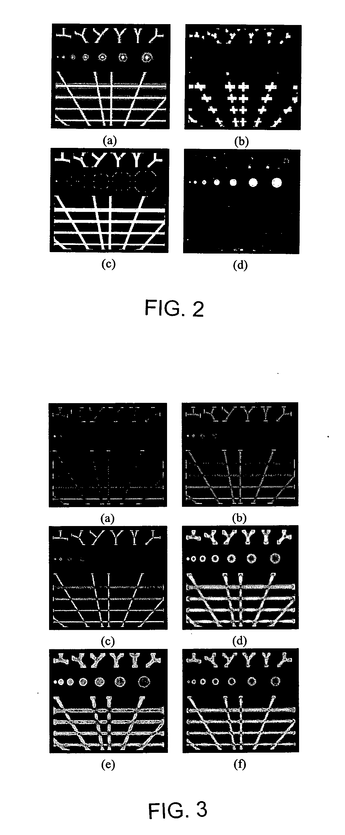
[0013] The invention still further comprehends a method for improving a diagnostic image including obtaining a thoracic diagnostic image, removing non-lung regions in the image to provide a lung image, enhancing vessels of a lung in the lung image according to a first-order partial derivative of each of a plurality of voxels of the lung image, constructing a vessel tree representation from the enhanced vessels, and subtracting the vessel tree representation from the lung image to enhance the visibility of nodules.
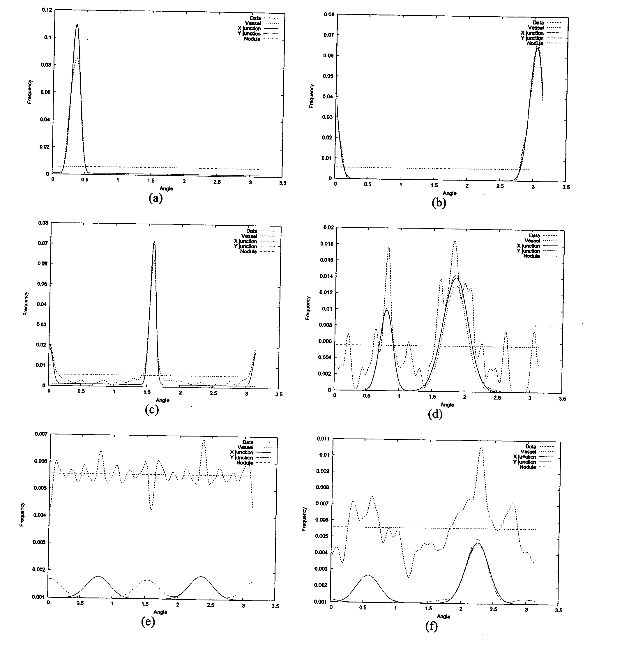
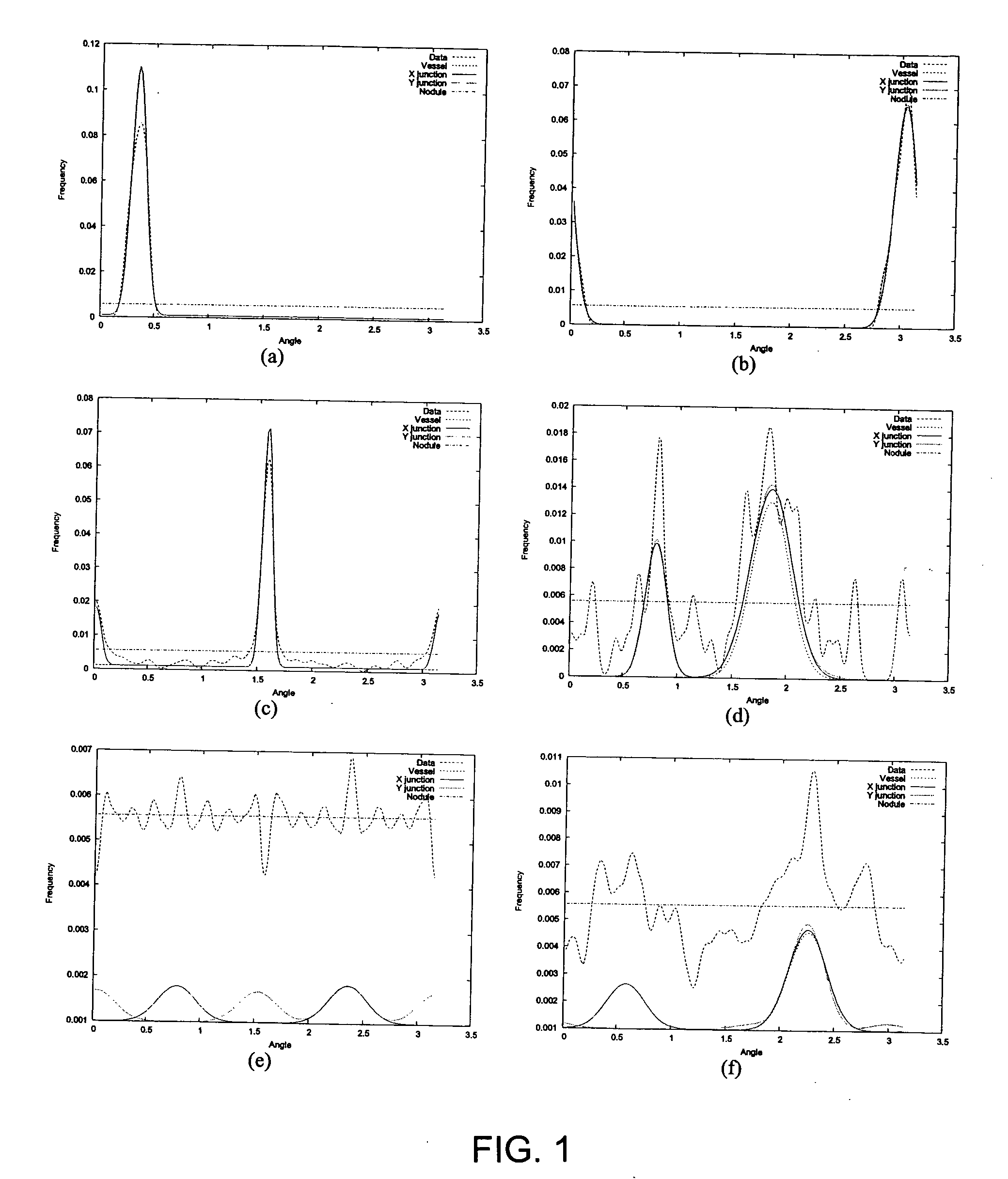
[0016] The vessel tree representation construction according to one embodiment of this invention is based on a fuzzy shape representation of the filtered or enhanced data, which is obtained by using regulated morphological operations. The construction of vessel tree representations, or “vessel trees,” begins by extracting vessel segments based on principal directions in the correlation matrix and collinearity constraints. These vessel segments are then merged into vessel trees. The algorithm may accommodate vessel bifurcation and discontinuities due to noise. The method of this invention reduces or eliminates false positives, such as are often generated by currently used nodule detection algorithms.

Problems solved by technology

The result is a large increase in the amount of medical imaging data that must be analyzed.
Manual analysis is often labor intensive and error prone.
Automated detection of lung nodules in thoracic CT scans is, for example, an important clinical challenge.
Manual analysis by a radiologist is generally time consuming, and may result in missed nodules.
Consequently, such small and disconnected segments of blood vessels are often classified erroneously as nodules.
Segmenting the image data correctly to distinguish between the tissue, nodules, and vessels is generally a difficult problem that has direct consequences for subsequent processing steps.
Incorrect segmentation can divide structures that should be connected or connect structures that should be separated, thus leading to incorrect interpretation of the data.
The estimation of the Hessian of the intensity function involves second-order partial derivatives and so is highly sensitive to noise.
Due to noise and smoothing, junctions are characterized by a high ratio of eigenvalues and so tend to be suppressed by vessel enhancement filters; this, in turn, leads to discontinuity of blood vessels.

Method used


Image

  • Method for enhancing diagnostic images using vessel reconstruction
  • Method for enhancing diagnostic images using vessel reconstruction
  • Method for enhancing diagnostic images using vessel reconstruction

Examples


Embodiment Construction

[0029] The present invention provides a method for improving a diagnostic image obtained by available imaging techniques, such as x-ray, magnetic resonance imaging (MRI), and computed tomography (CT). While the method of the invention is useful in improving various diagnostic images of any portion of the body, the invention will be described below with reference to a thoracic diagnostic image for imaging of the lungs by CT scanning.

[0030] In one embodiment of this invention, a diagnostic image is obtained by an available imaging technique, such as CT scanning. The diagnostic image generally is used to image a targeted object or portion of the body, and thus non-relevant regions, i.e., the portions of the obtained image that are not related to the desired or targeted object(s), body portion(s), or organ(s) are desirable removed. The non-relevant regions, e.g., non-lung regions of a thoracic image, can be removed by various and alternative means available to those skilled in the art....


PUM


Abstract

A method for improving a thoracic diagnostic image for the detection of nodules. Non-lung regions are removed from the diagnostic image to provide a lung image. Vessels and vessel junctions of the lung(s) in the lung image are enhanced according to a first-order partial derivative of each of a plurality of voxels of the lung image. A vessel tree representation is constructed from the enhanced vessels and vessel junctions. The vessel tree representation can be subtracted from the lung image to enhance the visibility of nodules in the lung(s).

Description

BACKGROUND OF THE INVENTION [0001] This invention is directed to a method for improving a diagnostic image, such as obtained by, for example, magnetic resonance imaging (MRI) or computed tomography (CT). [0002] Research in computer-aided diagnosis (CAD) has been facilitated by the rapid evolution in computer and medical imaging technologies in recent years. Three-dimensional diagnostic imaging tools, such as magnetic resonance imaging (MRI) and computed tomography (CT), are widely used for a vast range of applications. The result is a large increase in the amount of medical imaging data that must be analyzed. Manual analysis is often labor intensive and error prone. [0003] Automated detection of lung nodules in thoracic CT scans is, for example, an important clinical challenge. Manual analysis by a radiologist is generally time consuming, and may result in missed nodules. Furthermore, the amount of image data that has to be analyzed continues to increase. Blood vessel segmentation i...

Claims


Application Information

Patent Timeline
Patent Type & Authority Applications(United States)
IPC IPC(8): G06K9/00
CPCG06T7/0012G06T2207/30061G06T2207/30101G06T5/003G06T2207/20036G06T5/20G06T2200/04G06T2207/10081G06T5/005G06T5/77G06T5/73
Inventor AGAM, GADWU, CHANGHUA
Owner ILLINOIS INSTITUTE OF TECHNOLOGY
Learn More