
Acoustically induced blood stasis and in vivo optical spectroscopy

a technology of optical spectroscopy and acoustically induced blood stasis, which is applied in the field of therapeutic and in vivo diagnostic techniques, can solve the problems of blood cell banding in plasma medium, difficulty in measuring blood flow alterations, and no one has investigated diagnostic potential, etc., and achieve the effect of suppressing blood flow

Active Publication Date: 2007-03-08
UNIVERSITY OF ROCHESTER
View PDF10 Cites 7 Cited by
  • Summary
  • Abstract
  • Description
  • Claims
  • Application Information

AI Technical Summary

Benefits of technology

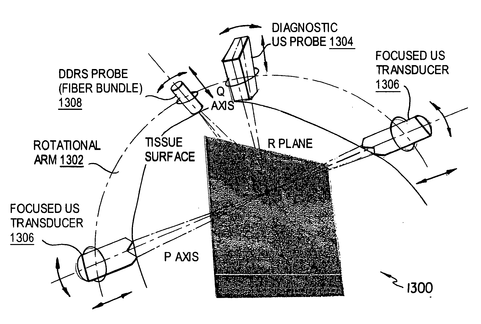
[0025] Temperature monitoring in the presence of blood flow and tissue heterogeneity is a known challenge in HIFU. We employ a simple solution for the breast by inserting tiny temperature measuring semiconductor probes (thermistors) interstitially in and around the mass to be ablated. Digital readout and feedback control are possible to ensure adequate but safe ablation power.
[0043] 9. Radiation beam is switched off; focal ultrasound is switched on to oxygenate the tumor mass; allow sufficient time for oxygenation of tumor mass.

Problems solved by technology

Even a traveling pressure wave, with small amounts of reflection at the tissue boundaries, can cause banding of blood cells in the plasma medium.
Many have continued to study the diagnostic limits and dangers of ultrasound and ultrasound-induced stasis, but to the best of our knowledge, no one has investigated the diagnostic potential.
A limiting factor in studying this ultrasound-induced phenomenon has been the difficulty of measuring the blood flow alterations.
The elevated temperatures are maintained for varying periods and frequencies, and cause cell death only in regions where the ultrasound is focused.
This technique inhibits the treatment of tumors near the chest wall and is dangerous because of the exposure of the chest cavity to high intensity ultrasound.

Method used


Image

  • Acoustically induced blood stasis and in vivo optical spectroscopy
  • Acoustically induced blood stasis and in vivo optical spectroscopy
  • Acoustically induced blood stasis and in vivo optical spectroscopy

Examples


Embodiment Construction

[0084] Preferred embodiments of the present invention will be set forth in detail with reference to the drawings, in which like reference numerals refer to like elements or steps throughout.

[0085]FIG. 1 shows an apparatus 100 according to the first preferred embodiment. The apparatus 100 includes a heater 102, a rubber absorber 104, an optical probe 106, a metal reflector 108, a Plexiglas restraint 110 for restraining a test subject (mouse) M, an ultrasound transducer 112, a positioning arm 114, and a Plexiglas water tank 116.

[0086] Ultrasound was generated by a ≈1 MHz piezoelectric ceramic crystal (Channel Industries) mounted behind a concave aluminum lens with a focal length of 7 cm. At 1 MHz the −6 dB focal zone diameter was 2 mm and the focal zone length was 30 mm. The ultrasound signal was created by a function generator (Agilent 33250A) and amplified by an RF amplifier (Amplifier Research 25A250A), monitored and recorded by an oscilloscope (Tektronix TDS 2022). The ultrasoni...


PUM


Abstract

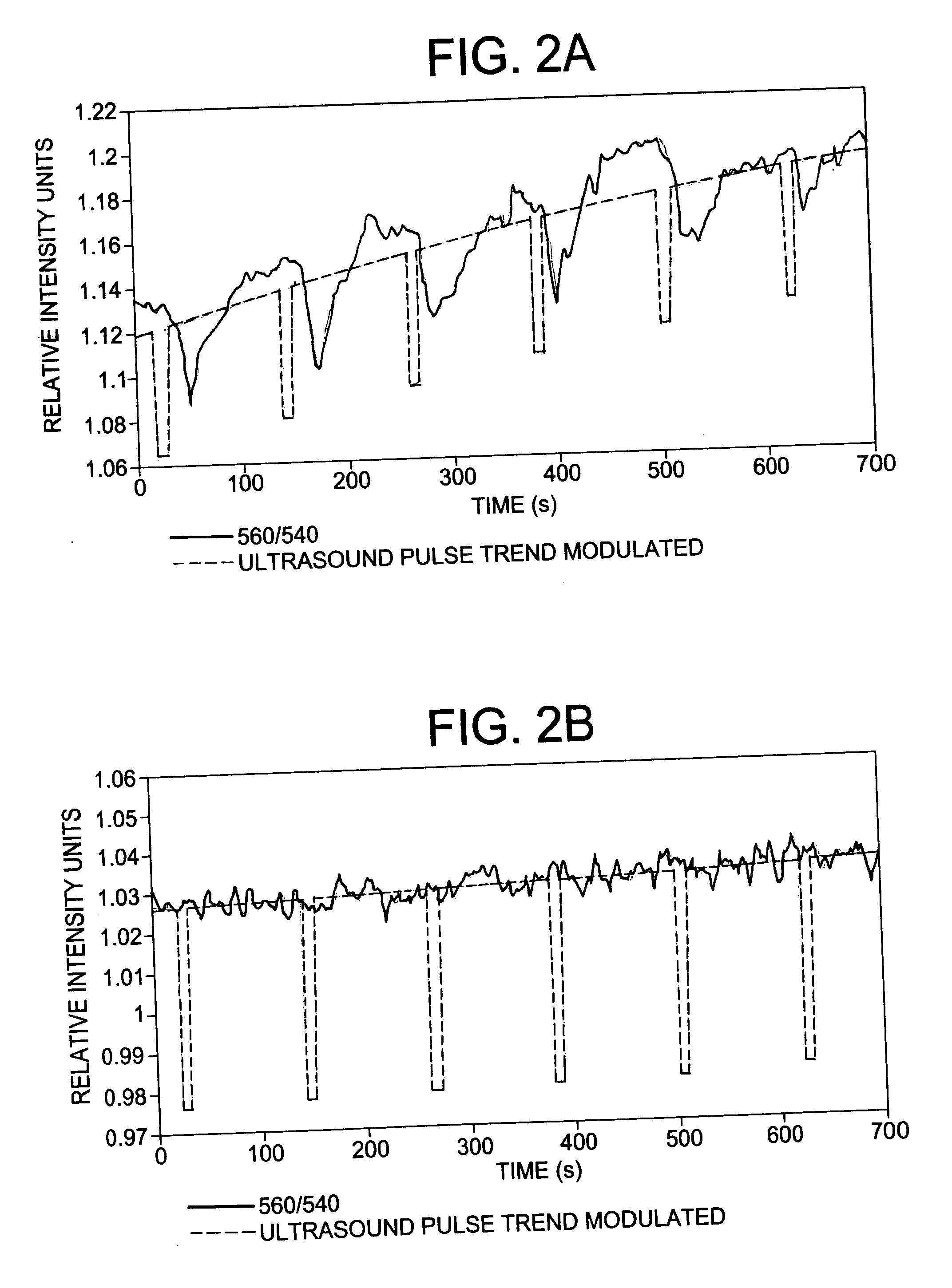
Ultrasound-induced blood stasis has been observed for more than thirty years. Most of the literature has been focused on the health risks associated with this phenomenon and methods employed to prevent stasis from occurring during ultrasound imaging. To date, experimental observations have been either in vitro or invasive. The current work demonstrates ultrasound-induced blood stasis in murine tumor and nontumor tissue, observed through noninvasive measurements of optical spectroscopy, and discusses possible diagnostic uses for this previously undesirable effect of ultrasound.

Description

REFERENCE TO RELATED APPLICATION [0001] The present application claims the benefit of U.S. Provisional Patent Application No. 60 / 709,108, filed Aug. 18, 2005, whose disclosure is hereby incorporated by reference in its entirety into the present disclosure.STATEMENT OF GOVERNMENT INTEREST [0002] The work leading to the present invention was supported by National Cancer Institute Grant No. CA107860. The government has certain rights in the invention.FIELD OF THE INVENTION [0003] The present invention is directed to therapeutic and diagnostic techniques and more particularly to therapeutic and in vivo diagnostic techniques combining acoustically induced blood stasis with optical spectroscopy. DESCRIPTION OF RELATED ART [0004] Stationary sound waves have long been known to create banding effects when solids are suspended in liquids; sand in air (in a cylinder), bubbles in water, etc. Stationary ultrasound waves can create bands of red blood cells in vitro in chick embryos removed from t...

Claims


Application Information

Patent Timeline
IPC IPC(8): A61B5/05A61B6/00
CPCA61B5/0075A61B5/0086A61N7/00A61B8/0825A61B8/12A61B5/4381
Inventor YU, YANWINEY, BRIANLIAO, LYDIA
Owner UNIVERSITY OF ROCHESTER
Learn More