Detection of rho proteins

a technology of rho proteins and detection methods, applied in the field of detection of rho proteins, can solve the problems of poor reproducibility of assays, low sensitivity, and inability to meet high throughput applications

Active Publication Date: 2007-01-25
CYTOSKELETON INC
View PDF5 Cites 9 Cited by
  • Summary
  • Abstract
  • Description
  • Claims
  • Application Information

AI Technical Summary

Benefits of technology

[0104] There are some sequence motifs that are shared between subgroups of Rho GTPase effectors, for example, the Cdc42 / Rac-interacting binding (CRIB) motif is present in many, though not all Rac and Cdc42 binding proteins (Burbelo et al., 1995, J. Biol. Chem., 270: 29071-29074) and has been found to be necessary but not sufficient for effector binding (Rudolph et al., 1998, J. Biol. Chem., 273: 18067-18076). A further common Rho effector motif is the Rho effector homology (REM) found in PRK1 and PRK2 and the HR1 repeat motif which is found in effectors such as rhophilin and rhotekin (Flynn et al., 1998, J. Biol. Chem., 273: 2698-2705). However, there are a large number of effectors that do not contain any of the currently identified GTPase-binding motif / s. These include the POSH, P13K and DAG effectors (Bishop et al., 2000, Biochem. J., 348: 241-255). This type of protein is classified as a Rho effector purely on its functional ability to differentiate between the GTP (active) and GDP (inactive) bound forms of Rho GTPases (Tapon, 1998, EMBO J., 17: 1395-1404; and Kobayashi et al., 1998, J. Biol. Chem., 273: 291-295). It is therefore accepted by those skilled in the art that Rho effectors are currently defined by their functional ability to selectively recognize the GTP-bound (active) form of Rho GTPases over the inactive GDP form of the GTPase (Vetter et al., 2001, Science, 294: 1299-1304; Blumenstein et al., 2004, J. Biol. Chem., 279: 53419-53426; Martin et al., 1995, EMBO J., 14: 1970-1978; Leung et al., 2005, Proc. Natl. Acad. Sci., 102: 5685-5690; Bishop et al., 2000, Biochem. J., 348: 241-255; and Fujisawa et al., 1998, J. Biol. Chem., 273: 18943-18949).
[0105] The example provided herein has utilized six of the most extensively characterized Rho effectors, or more specifically Rho effector-GTPase Binding Domain (GBD) peptides. Table 1 details the effector and the specificity of the effector for activated Rho family proteins. TABLE 1Effector Recognition of Activated Rho Family ProteinsActivated Rho familyEffectorproteins recognizednameby effector-GBDReferencesRhotekinRhoA, RhoB, RhoCReid, 1996, J. Biol. Chem.,271: 13556ROCK1RhoA, RhoB, RhoC,Fujisawa, 1996, J. Biol. Chem.,RhoE / Rnd3271: 23022Riento, 2003, Mol. Cell. Biol.,23: 4219PAK1Rac1, Rac1b, Rac2,Bagrodia, 1995, J. Biol. Chem.,Rac3, Cdc42, Wrch1270: 22731Saras, 2004, J. Exp. Cell Res.,299: 356POSHRac1Tapon, 1998, EMBO, 17: 1395WASPCdc42, TC10, TCL,Symons, 1996, Cell, 84: 723Wrch2 / ChpAbe, 2002, J. Cell. Sci., 116:155Vignal, 2000, J. Biol. Chem.,275: 36457Aronheim, 1998, Curr. Biol.,8: 1125Dia1RhoA, RhoCWatanabe, 1997, EMBO J., 16:30444
[0106] For all of the effector proteins and effector GTPase Binding Domains (GBD) provided herein as examples, the full length mammalian cDNA has been previously described (see Table 2). Effector-GBD peptides have been cloned by PCR using the primers and cDNA sources indicated in Table 2. The nucleotide numbering in Table 2 corresponds to the Genbank submission numbering scheme. GTPase Binding Domains (GBDs) were selected based upon published data identifying active Rho binding domains of effectors (see Table 1). TABLE 2Effector Cloning InformationNucleotidesEffectorAccessionPrimers for cDNA cloningincludedcDNAnamenumber5′ primer 3′ primer(amino acid)sourceRhotekinNM_0091065′ caggatccattctggaagatctgaacatgctg19-267Mouse(SEQ ID NO: 98)(7-89)3′ cagaattcgcccccaccagttttttcgag(SEQ ID NO: 99)ROCK1-1NM_0054065′ accgaattcgaagctgagcaatatttctcg2539-3070 Human(SEQ ID NO: 100)(847-1024)3′ accgaattctcaacattgtgtattagctttctttctata(SEQ ID NO: 101)Dia1-1NM_0052195′ caggatcctctgcatcatatggggatgat187-780 Human(SEQ ID NO: 102)(63-260)3′ cagaattctcatagaatacaaagagcagaaag(SEQ ID NO: 103)PAK1NM_0025765′ caggatccaaagagaaagagcggccagagat202-453 Human(SEQ ID NO: 104)(68-151)3′ cagaattctcaacactcagctgacttatctgtaaagctc(SEQ ID NO: 105)POSHNM_0208705′ caggatccaagcaccccgacaccaagaag871-1089Human(SEQ ID NO: 106)(291-363) 3′ cagaattcacccagtggtgcttatatggacc(SEQ ID NO: 107)WASPNM_0003775′caggatccgacatccagaaccctgacatcacg601-963 Human(SEQ ID NO: 108)(201-321) 3′cagaattctcaacatcgagatggcggtgggggcggc(SEQ ID NO: 109)
[0107] The sequences from the GenBank accession numbers from Table 2 and anywhere provided herein, are incorporated herein by reference in their entirety.
[0108] In instances where effector-GBD peptides were linked covalently to maleimide plates (see later in this example and Table 3), the effector-GBD peptide DNA was modified to contain a single cysteine residue at the carboxy terminal. Cysteine codons were engineered into the primer design for ROCK1, PAK1 and WASP (see Table 2, the cysteine codon in the 3′ primers are underlined). In the case of POSH, advantage of the cystein codon (position 351) close to the carboxy terminal was taken.
[0109] The effector-GBD of rhotekin contains three internal cysteine residues (Table 2 and FIG. 1). To allow directional binding of rhotekin-GBD to maleimide plates the rhotekin-GBD was chemically synthesized (Entelechon GmbH, St. Veit-Weg 2, 93051 Regensburg, Germany), the three internal cysteins were removed and replaced with glutamic acid (aa# 43), leucine (aa# 49) and serine (aa# 65) respectively. A cysteine codon was placed at the carboxy terminal of the modified rhotekin-GBD, immediately upstream of a stop codon. The design of the synthetic rhotekin-GBD sequence was carried out using Leto 1.0 gene 20 optimization software which is based on a proprietary genetic algorithm (Entelechon GmbH, St. Veit-Weg 2, 93051 Regensburg, Germany). The sequence design allowed optimization of codon usage, homogenous GC content, mRNA secondary structure, codon and motif repeats and restriction sites. The synthetic Rhotekin-GBD DNA was supplied by Entelechon GmbH in a pUC18 cloning vector. The DNA was cleaved with restriction enzymes BamH1 and EcoR1 according to the manufacturers instructions (Promega Corp., Madison, Wis.) and the 273 bp DNA fragment containing modified Rhotekin-GBD was cloned directly into the BamH1 and EcoR1 sites of the expression vector pGEX-4T, creating a Rhotekin-GBD domain fused to a Glutathione S Transferase tag at the amino terminal and containing a unique cysteine residue at the carboxy terminal. All molecular biology manipulations were carried out in accordance with general principles outlined in Molecular Cloning, A Laboratory Manual, 1998, Second edition, Cold Spring Harbor Laboratory Press. Ed. Sambrook et al.

Problems solved by technology

The assay suffers from poor reproducibility due to the multiple manipulations required in the performance of the assay, and low sensitivity.
It is also not suitable for high throughput applications (Teusch et al., 2006, Assay and Drug Devel., 4: 133).
Because the placing of the environmental dye on any particular probe requires extensive analysis and a particular effector may not be compatible with dye linkage, the usefulness of any particular effector cannot be predicted (Nalbant et al., 2004, Science, 305: 1615-1619).
Furthermore, because the use of direct effector detection in vivo results in a probe that most often recognizes more than one GTPase, specificity is an issue in these assays.
A further issue with this type of assay is that introduction of exogenous effector will actually alter the levels of Rho GTPase activation, which creates a technically challenging assay method (Pertz et al., 2004, J. Cell Sci., 117: 1313-1318).
Furthermore, that the dyes respond to environmental changes poses issues to drug screening applications (Hahn et al., U.S. Pat. No. 6,951,947 B2).
Furthermore, since a Rho-specific antibody is not involved in this method, the specificity of the assay is limited.
This assay was developed to replace the GST pull-down assay due to its incompatibility for high throughput screening and poor reproducibility.
Since actin cytoskeleton is regulated by multiple signal pathways, the specificity for this assay is very limited.

Method used


Image

  • Detection of rho proteins
  • Detection of rho proteins
  • Detection of rho proteins

Examples


example 1

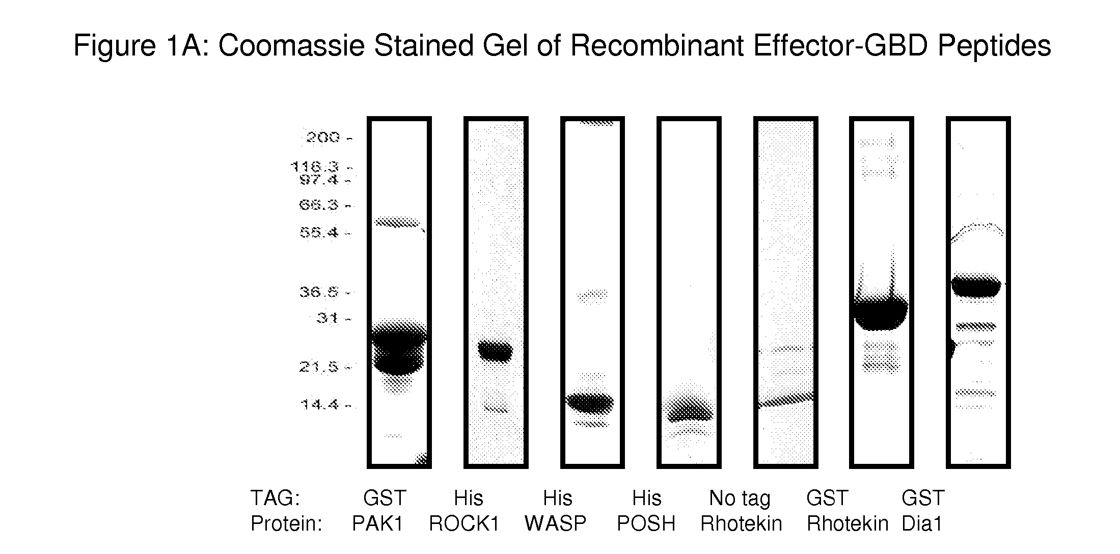
Production of Effector-GBD Peptides for the G-LISA Assay

[0104] There are some sequence motifs that are shared between subgroups of Rho GTPase effectors, for example, the Cdc42 / Rac-interacting binding (CRIB) motif is present in many, though not all Rac and Cdc42 binding proteins (Burbelo et al., 1995, J. Biol. Chem., 270: 29071-29074) and has been found to be necessary but not sufficient for effector binding (Rudolph et al., 1998, J. Biol. Chem., 273: 18067-18076). A further common Rho effector motif is the Rho effector homology (REM) found in PRK1 and PRK2 and the HR1 repeat motif which is found in effectors such as rhophilin and rhotekin (Flynn et al., 1998, J. Biol. Chem., 273: 2698-2705). However, there are a large number of effectors that do not contain any of the currently identified GTPase-binding motif / s. These include the POSH, P13K and DAG effectors (Bishop et al., 2000, Biochem. J., 348: 241-255). This type of protein is classified as a Rho effector purely on its functio...

example 2

Utility of Covalently Linked Effector-GBD Plates in G-LISA Assays

[0124] There are many chemistries described in the art for linking a particular peptide to a microtiter plate or microarray (Dent et al., 1998, Bioconjugation, Macmillan Reference Ltd, Chapter 8: 505-556). In the broadest terms, linkages can be classified into covalent and non-covalent formats. Both types of linkage are demonstrated herein (see non-covalent linkage, Example 7). The example described herein details a method of covalent linkage through maleimide activated plates.

[0125] Covalent linkage of peptides to microtiter plates through activated surface groups occurs when electrons are shared between atoms to generate chemical bonds. While the reactions forming such bonds are always theoretically reversible, the low probability of the reverse reaction occurring in practice results in products that can be thought of as permanent (Dent et al., Bioconjugation, Macmillan Reference Ltd, 1998, p. 218-342). The essent...

example 3

Development of Antigen Presenting Buffer for the G-LISA

[0144] Initial attempts to detect active Rho GTPases in the RhoA: ROCK and Rac1 : POSH G-LISA assays were not successful because a signal that was significantly above background in either assays (FIGS. 5A (untreated) and 5B (no APB)) could not be obtained. An initial strategy was to develop an ELISA based assay by following a protocol essentially analogous to the original pull-down assays described by Benard et al. for the Rac1: PAK pull-down assay (1999, J. Biol. Chem., 274: 13198-13204) and Ren et al. and Kranenburg at al. for the RhoA: Rhotekin pull-down assay (1999, EMBO J., 18: 578; and 1999, Mol. Biol. Cell, 10: 1851-1857, respectively). The same procedure has been used for pull-down assays using ROCK, Citron, Dia and WASP (Kimura et al., 2000, J. Biol. Chem., 275: 17233-17236, and Edlund et al., 2002, Mol. Biol. Cell, 13: 902-914). Briefly, maleimide plates were coated with effector, active Rho GTPases in standard pull-...


PUM

PropertyMeasurementUnit
pHaaaaaaaaaa
concentrationaaaaaaaaaa
affinityaaaaaaaaaa

Abstract

The present invention provides methods of detecting activated Rho GTPase proteins by contacting a solid support with a sample comprising an activated Rho GTPase protein. The solid support is linked to an activated Rho GTPase binding peptide. The activated Rho GTPase protein remains associated with the solid support during the detection.

Description

CROSS-REFERENCE TO RELATED APPLICATIONS [0001] This application claims priority to U.S. Provisional Application Ser. No. 60 / 695,844, filed Jul. 5, 2005 which is incorporated herein by reference in its entirety.FIELD OF THE INVENTION [0002] The present invention is directed, in part, to methods of detecting activated Rho proteins. BACKGROUND OF THE INVENTION [0003] The Rho GTPases are a structurally and functionally distinct GTPase of the Ras superfamily (Wennerberg et al., 2005, J. Cell Sci., 118: 843-846; Takai et al., 2001, Physiol. Rev., 81: 153-208). They are involved in a diverse array of cellular functions, including the regulation of actin and tubulin dynamics, cell polarity, membrane transport pathways and transcription factor activities (Ridley et al., 1992, Cell, 70: 389-399; Braga et al., 1997, J. Cell Biol., 137: 1421-1431; and Coso et al., 1995, Cell, 81: 1137-1146). However, some members of the family (e.g., RhoBTB1, RhoBTB2 and RhoH) do not appear to be functionally c...

Claims


Application Information

Patent Timeline
Patent Type & Authority Applications(United States)
IPC IPC(8): G01N33/53
CPCG01N2333/916G01N33/573
Inventor CHENG, LIDAVIS, ASHLEYMIDDLETON, KIM
Owner CYTOSKELETON INC
Why Eureka
Try Eureka