Eureka AIR delivers breakthrough ideas for toughest innovation challenges, trusted by R&D personnel around the world.

Structure of a glucocorticoid receptor ligand binding domain comprising an expanded binding pocket and methods employing same

a glucocorticoid receptor and ligand binding technology, applied in the field of glucocorticoid receptor polypeptides, can solve the problems of inability to use crystals in the ligand binding domain of human glucocorticoid, inability to accurately represent the structure of x-rays, and models that have some utility

Inactive Publication Date: 2007-01-25
SMITHKLINE BECKMAN CORP
View PDF1 Cites 23 Cited by
  • Summary
  • Abstract
  • Description
  • Claims
  • Application Information

AI Technical Summary

Benefits of technology

[0021] A crystalline GR polypeptide complex comprising an expanded binding pocket is disclosed. Preferably, the crystalline form has lattice constants of of a=b=127.656 Å, c=87.725 Å, α=90°, β=90°, γ=120°. Preferably, the crystalline form is a hexagonal crystalline form. More preferably, the crystalline

Problems solved by technology

Unfortunately, such crystals have been unavailable for the ligand binding domain of a human glucocorticoid receptor, as well as many other proteins of interest.
These theoretical models have some utility, but cannot be as accurate as a true X-ray structure, such as the X-ray structure disclosed here.
Because of their limited accuracy, a model for GRα will generally be less useful than an X-ray structure for the design of agonists, antagonists and modulators of GRα.
Potential or existent homology models or existing crystal structures cannot provide the necessary degree of specificity.
Still other journal articles have reported E.coli expression of GST-GR, but also noted a failure to purify the purported polypeptide.
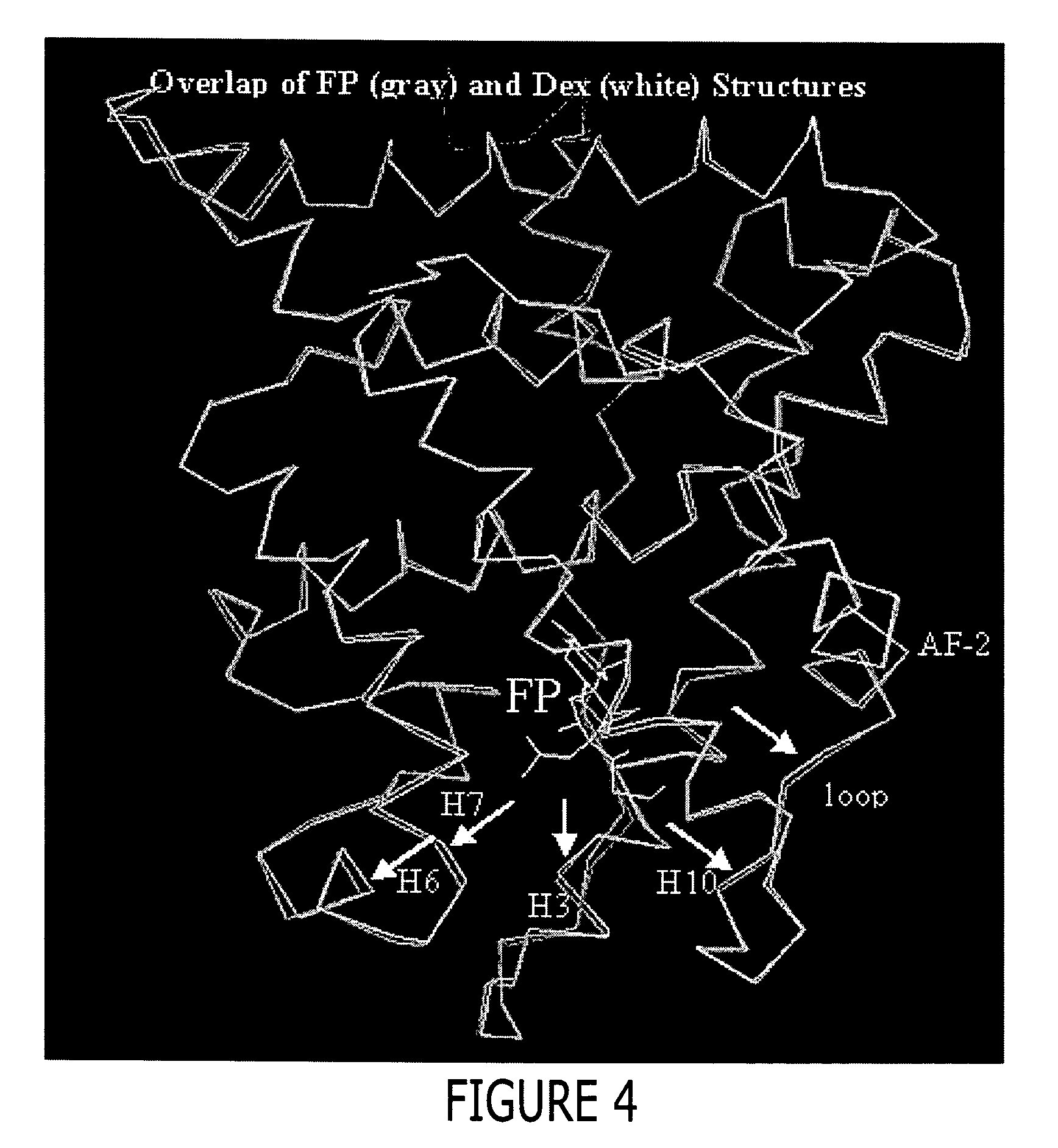
While offering unprecedented insight into the structure of GR in complex with a ligand, this structure does not adequately answer the question surrounding the higher affinity of GR for FP than for dexamethasone.
Nor can available GR, AR, MR and PR models adequately explain the mode of FP association with these NRs.
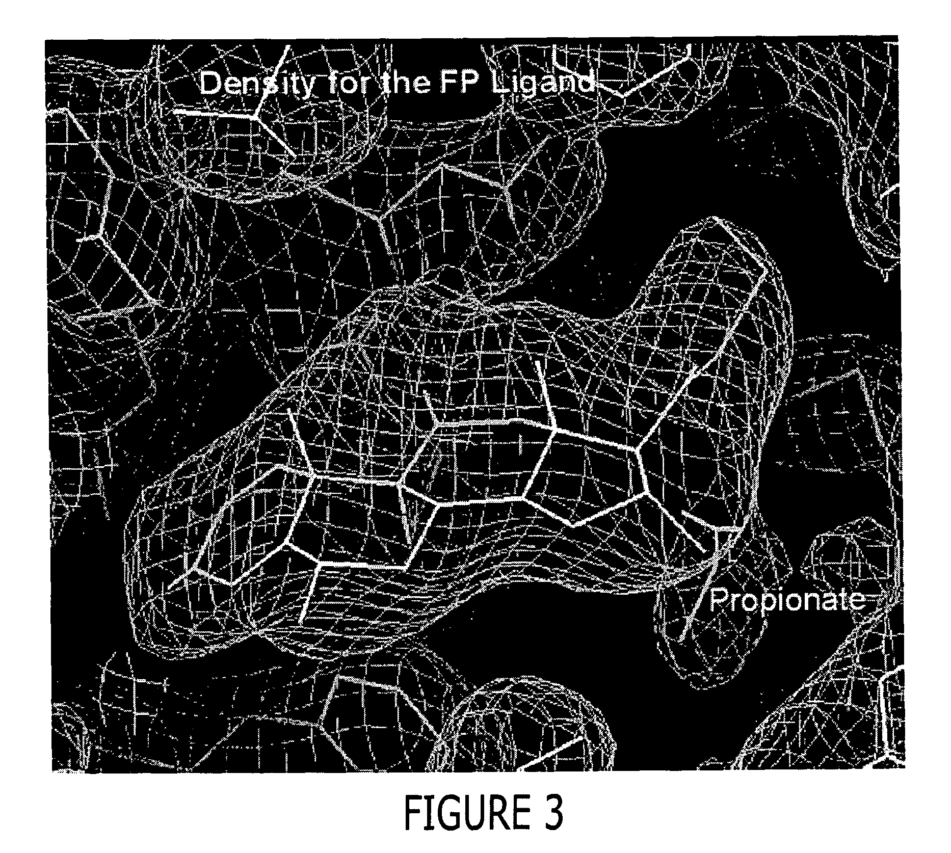
Examination of these models indicates that the ligand binding pocket is sterically limited in its ability to accommodate FP and other ligands, such as steroidal molecules having large substituents at the C-17α position and non-steroidal molecules having substituents predicted to fill the same space as would be filled by the proprionate group of FP.

Method used


Image

  • Structure of a glucocorticoid receptor ligand binding domain comprising an expanded binding pocket and methods employing same
  • Structure of a glucocorticoid receptor ligand binding domain comprising an expanded binding pocket and methods employing same
  • Structure of a glucocorticoid receptor ligand binding domain comprising an expanded binding pocket and methods employing same

Examples


example 1

Laboratory Example 1

Expression of a GRα Polypeptide

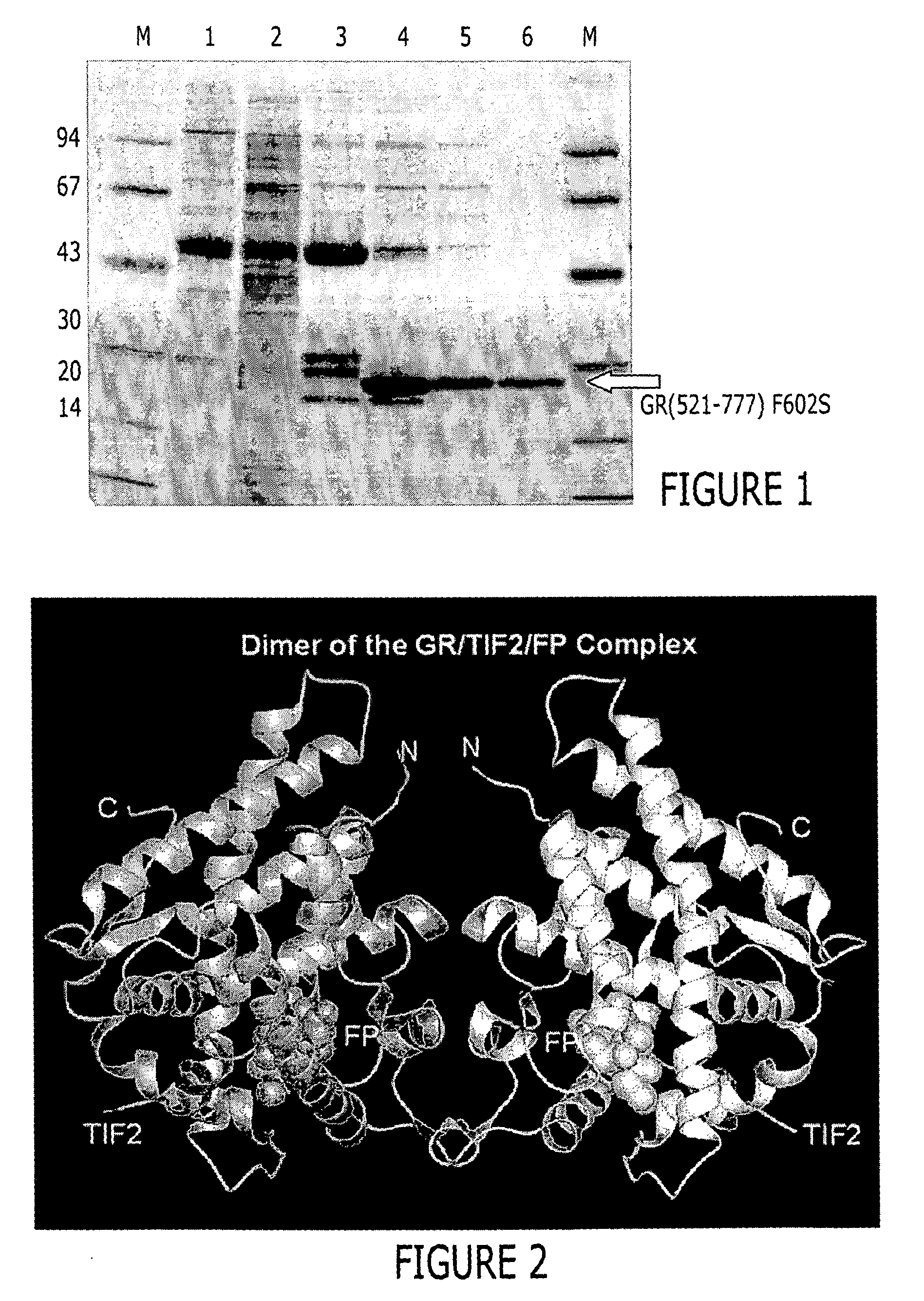
[0450] BL21(DE3) cells (Novagen / Invitrogen, Inc., Carlsbad, Calif., United States of America) were transformed with the expression plasmid 6xHisGS-TGR(521-777) F602S pET24 following established protocols. Following overnight incubation at 37° C. a single colony was used to inoculate a 10 ml LB culture containing 50 μg / ml kanamycin (Sigma, St. Louis, Missouri, United States of America). The culture was grown for ˜8 hrs at 30° C. and then a 500 μl aliquot was used to inoculate flasks containing 1 liter CIRCLE GROW™ media (Bio 101, Inc., Vista, Calif., United States of America) and the required antibiotic. The cells were then grown at 22° C. to an OD600 between 2 and 3 and then cooled to 18° C. Following a 30 min equilibration at that temperature, dexamethasone (Spectrum Chemical Co., Gardena, Calif., United States of America) (50 or 100 μM final concentration) was added. Induction of expression was achieved by adding IPTG (BACHEM, Ph...

example 2

Laboratory Example 2

Purification of a GR LBD (521-777) F602S Polypeptide Bound to Fluticasone Propionate

[0452] Approximately 37 g of cells were resuspended in 500 mL lysis buffer (50 mM Tris pH=8.0, 150 mM NaCl, 2M urea, and 30 μM fluticasone propionate) and lysed by passing 3 times through a Rannie APV Lab 2000 homogenizer (Rannie APV, Copenhagen, Denmark). The lysate was subjected to centrifugation (30 minutes, 20,000 g, 4° C.). The cleared supernatant was filtered through coarse pre-filters and 50 mM Tris, pH=8.0, containing 150 mM NaCl and 1M imidazole was added to obtain a final imidazole concentration of 50 mM. This lysate was loaded onto a XK-26 column (Pharmacia, Peapack, N.J.) packed with Sepharose [Ni2+ charged] chelation resin (Pharmacia, Peapack, N.J.) and pre-equilibrated with lysis buffer supplemented with 50 mM imidazole. Following loading, the column was washed to baseline absorbance with equilibration buffer. This was followed by a linear (0 to 10%) glycerol and (2...

example 3

Laboratory Example 3

Preparation of a GR / TIF2 / Fluticasone Proprionate (FP) Complex

[0454] The GR / TIF2 / FP complex was prepared by adding a 1.2-fold excess of a TIF2 peptide containing sequence of KENALLRYLLDKDD (SEQ ID NO: 9) during the buffer exchange step as described below. The above complex was concentrated then diluted 1:1 with a buffer containing 500 mM NH4OAC, 50 mMTris, pH 8.0, 10% glycerol, 10 mM dithiothreitol (DTT), 0.5mM EDTA and 0.05% β-octyl-glucoside and concentrated to 1 ml. The complex was diluted 1:9 with the above buffer and slowly concentrated to 7.5 mg / ml in the presence of an additional 1.2 fold excess of a TIF2 peptide (residues 740-753), aliquoted and stored at −80° C.


PUM

PropertyMeasurementUnit
Fractionaaaaaaaaaa
Fractionaaaaaaaaaa
Fractionaaaaaaaaaa

Abstract

A solved three-dimensional crystal structure of a glucocorticord receptor (GR) α ligand binding domain polypeptide is disclosed, in the form of a crystalline glucocorticord receptor α ligand binding domain polypeptide in complex with the ligand fluticasone propionate (FP) and a peptide derived from the co-activator TIF2. The GR / FP / TIF2 structure includes an expanded binding pocket not seen in other GR structures. Methods of designing steroid and non-steroid modulators of the biological activity of GR and other nuclear receptors (NRs) are also disclosed. In another aspect of the present invention homology models of androgen receptor (AR), progesterone receptor (PR) and mineralcorticoid receptor (MR) are disclosed, as well as methods of forming homology models for other NRs. Methods of forming a soluble GR / FP / TIF2 complex are also disclosed.

Description

TECHNICAL FIELD [0001] The present invention relates generally to a glucocorticoid receptor polypeptide, to a glucocorticoid receptor ligand binding domain polypeptide, and to the structure of a glucocorticoid receptor ligand binding domain bound to fluticasone propionate and a co-activator peptide. This stucture reveals an expanded binding pocket having a configuration and volume not observed in other GR structures, which explains the observed binding of some ligands to GR. In one aspect, the invention relates to methods by which a soluble complex comprising glucocorticoid ligand binding domain, fluticasone propionate and a co-activator can be generated. Methods by which modulators and ligands of nuclear receptors, particularly steroid receptors, and more particularly glucosteroid receptors, and the ligand binding domains thereof, can be identified are also disclosed. The invention further relates to homology models of nuclear receptors, preferably the ligand binding domains of nuc...

Claims


Application Information

Patent Timeline
IPC IPC(8): G01N33/53G06F19/00G01N33/48G01N33/50C07K14/705A61K38/00C07K14/72
CPCA61K38/00G01N2333/723C07K2299/00C07K14/721
Inventor BLEDSOE, RANDY K.LAMBERT, MILLARD H. IIIMONTANA, VALERIE G.STEWART, EUGENE L.XU, H. ERIC
Owner SMITHKLINE BECKMAN CORP
Why Eureka
Eureka Blog
Learn More