
Use of neuropeptides for ligament, cartilage, and bone healing

a neuropeptide and ligament technology, applied in the direction of gastrin/cholecystokinin, prosthesis, metabolism disorder, etc., can solve the problems of pain, inflammation, instability of the affected joint, progressive degeneration of the joint or associated organ, and increase discomfort and difficulty of use, so as to speed up the healing of subchondral bone injuries and defects, accelerate the time to complete recovery, and promote the healing of damaged areas

Inactive Publication Date: 2006-11-02
WISCONSIN ALUMNI RES FOUND
View PDF3 Cites 69 Cited by
  • Summary
  • Abstract
  • Description
  • Claims
  • Application Information

AI Technical Summary

Benefits of technology

The present invention provides methods and pharmaceutical compositions for promoting healing and improving the functional properties of damaged or diseased connective tissues, such as ligaments, cartilage, and bone. The methods involve administering neuropeptides to the tissue to induce repair and improve the healing process. The pharmaceutical compositions contain a combination of a neuropeptide and a pharmaceutically suitable carrier. The invention is particularly useful for treating degenerative cartilage disease, traumatic cartilage injury, and osteochondral defects. It also has applications in promoting ligament and tendon healing and increasing the functionality of intact cartilage as a low-friction, load-bearing surface. The invention is also useful for speeding the healing of subchondral bone injuries and defects. Overall, the invention provides new methods and compositions for promoting connective tissue healing and improving their function.

Problems solved by technology

These disorders have many pathologic events in common and all result in pain, inflammation, and instability of the affected joint.
In many cases, connective tissue diseases can lead to progressive degeneration of the joint or associated organ with increasing discomfort and difficulty of use.
Moreover, peripheral neuropathies commonly associated with diabetes frequently manifest themselves as joint disease, poor tissue healing, and / or a compromised immune response.
Diabetic pathologies include defects in connective tissue, such as poor wound healing and diminished bone formation and growth.
While the causes of various diabetic neuropathies are not completely clear, it is thought that increased blood glucose levels over time result in damage to the blood vessels, connective tissue, nerves and other organs.
Further, it is unclear whether a single diabetic pathology promotes the pathological condition of another tissue, or whether each separate pathological condition independently results from increased blood glucose levels.
Peripheral nerve disorders are prevalent and can be debilitating, especially among diabetic populations.
This is particularly evident in the connective tissues of joints, including tendons, ligaments and cartilage, due to their constant use and visible loss of function.
In general, damaged nerves have limited potential for regeneration, and healing in joints following nerve damage is slow and often results in permanent joint damage.
Despite the prevalence of nerve disorders, little work has been done to elucidate how damage to nervous tissue leads to structural damage to various connective tissues.
However, continuing research suggests that these neurogenic compounds are produced throughout the nervous system and have complex effects.
The anterior cruciate ligament (ACL) has very little healing potential, while the medial cruciate ligament (MCL) has a greater healing potential (although certainly not ideal).
The scar tissue associated with ligament healing is mechanically inferior and compositionally abnormal as compared to uninjured tissue.
Healing after ligament injury can then result in ligament laxity, joint instability, prolonged pain, and abnormal joint motion.
Damage to sensory nerves can result in dysesthesia (perception of abnormal sensations), while damage to sympathetic nerves can result in wide spread organ problems due to alterations in blood flow (Association, 2000).
Substance P depletion by capsaicin has been shown to be detrimental to wound healing (Dunnick et al., 1996; Dwyer et al., 2003).
A decrease in immune cells (CD4+ and CD8+ cells) in whole blood has been shown to result from administering capsaicin (Santoni et al., 1996) to rats, which may account for poor healing of tissues following administration of capsaicin.
While techniques of inhibiting sensory information resulting from peripheral neuropathies affords relief from pain, these techniques do not treat the underlying cause of the pain, and in many instances may further potentiate the connective tissue degeneration that is the source of, or results from, pain induction.

Method used


Image

  • Use of neuropeptides for ligament, cartilage, and bone healing
  • Use of neuropeptides for ligament, cartilage, and bone healing
  • Use of neuropeptides for ligament, cartilage, and bone healing

Examples


example 1

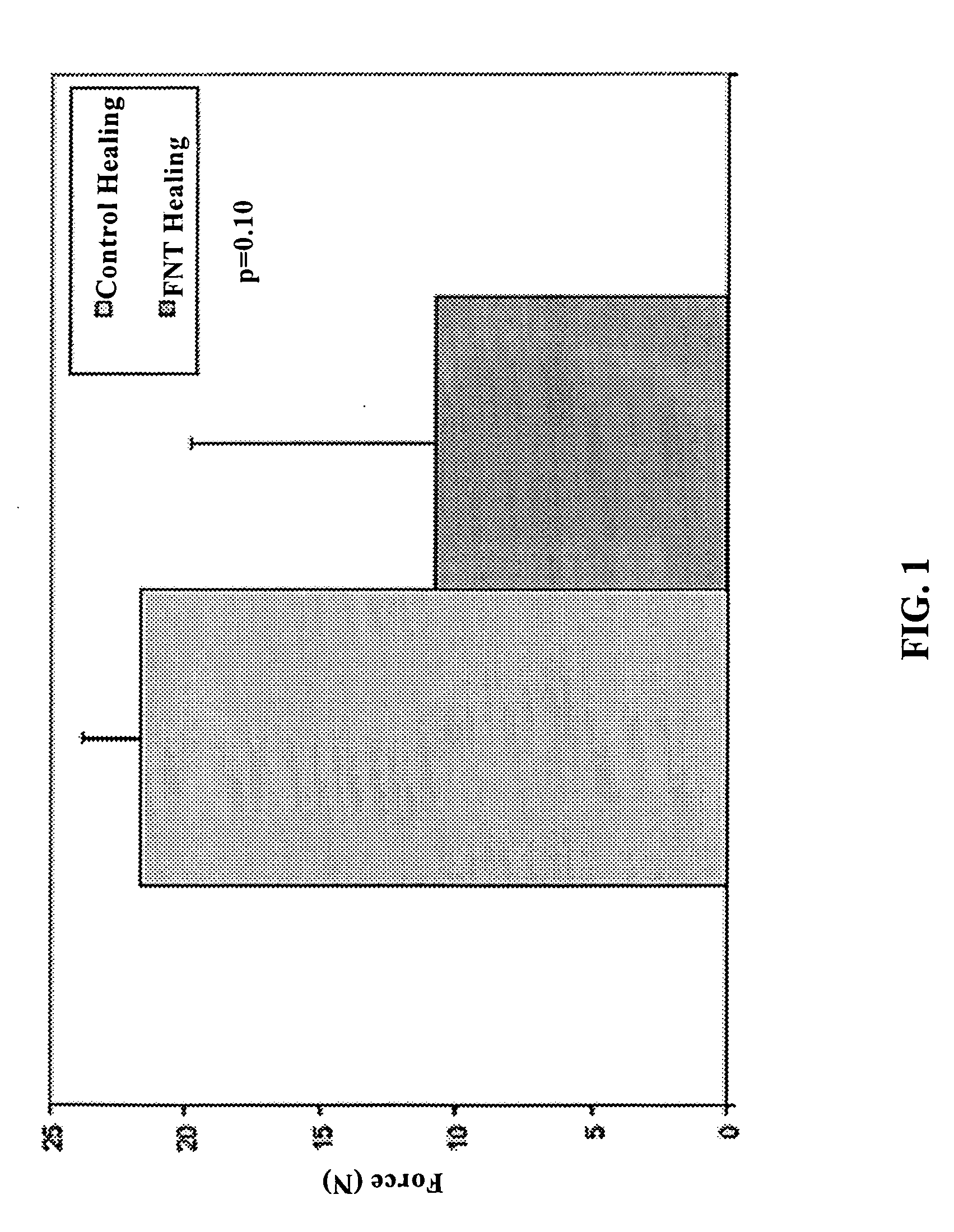
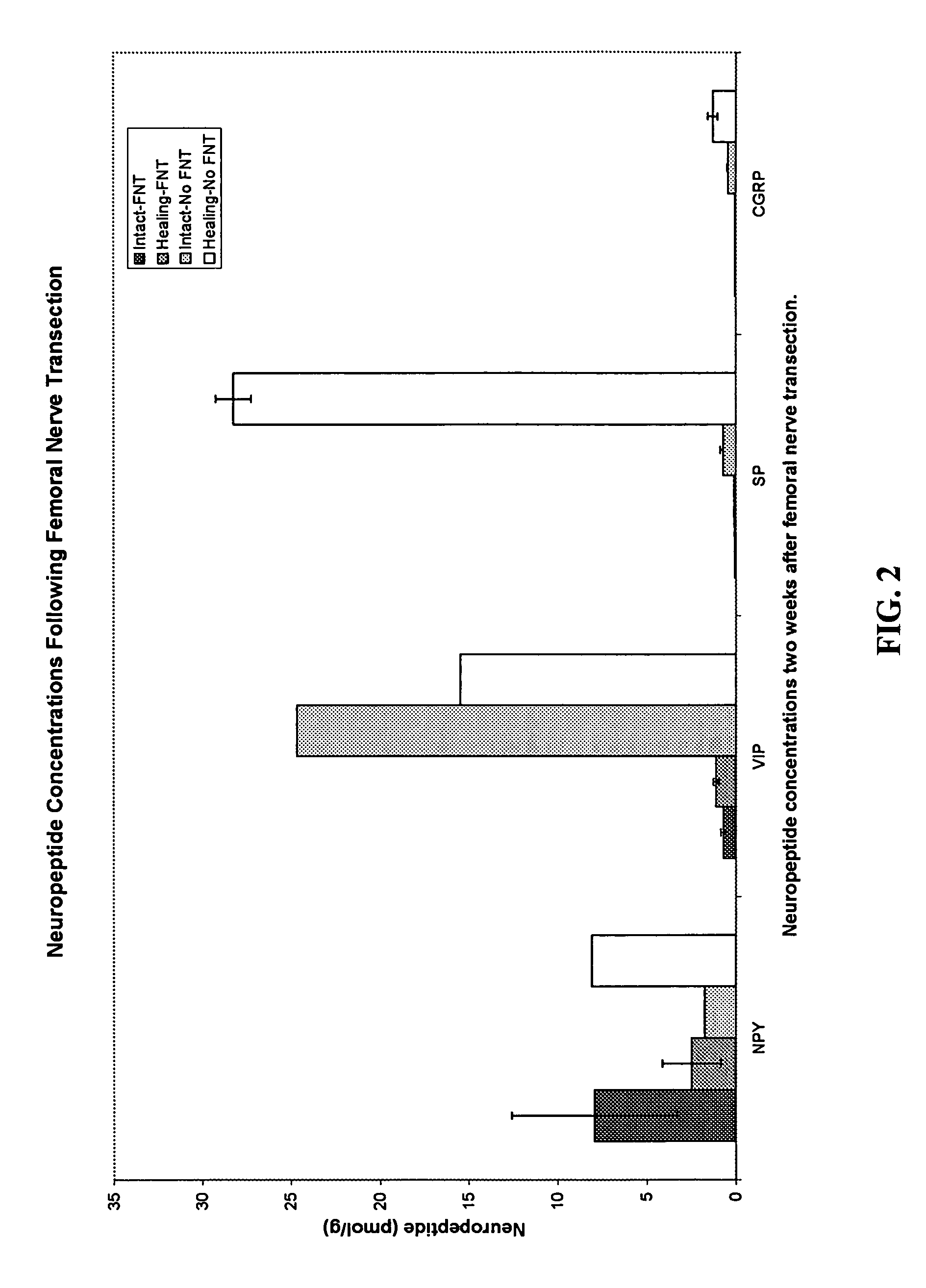
Femoral Nerve Transection Impairs Healing in a Rat MCL Model

[0076] As noted earlier, ligament healing is a complex problem that is influenced by inflammatory responses, extracellular matrix remodeling, and chemical mediators. The role of neurogenic factors in general, and neuropeptides in particular, in ligament healing remains undefined. The purpose of this Example was to investigate the role of sensory nerves on MCL healing in a neuropathic rat model.

[0077] Surgical Procedure: Thirteen female Wistar rats (278 g to 315 g) were divided into two groups: femoral nerve transection (FNT) or sham control. All rats were anesthetized with isofluorane (isofluorane 0.5-3%). Anesthesia was initiated in an induction chamber and maintained with a facemask. Skin covering the anterior inguinal region was shaved and prepared for surgery. A skin incision approximately 15 mm long was made and the fascia distal to the inguinal was incised. The femoral artery, vein, and nerve were exposed and the fe...

example 2

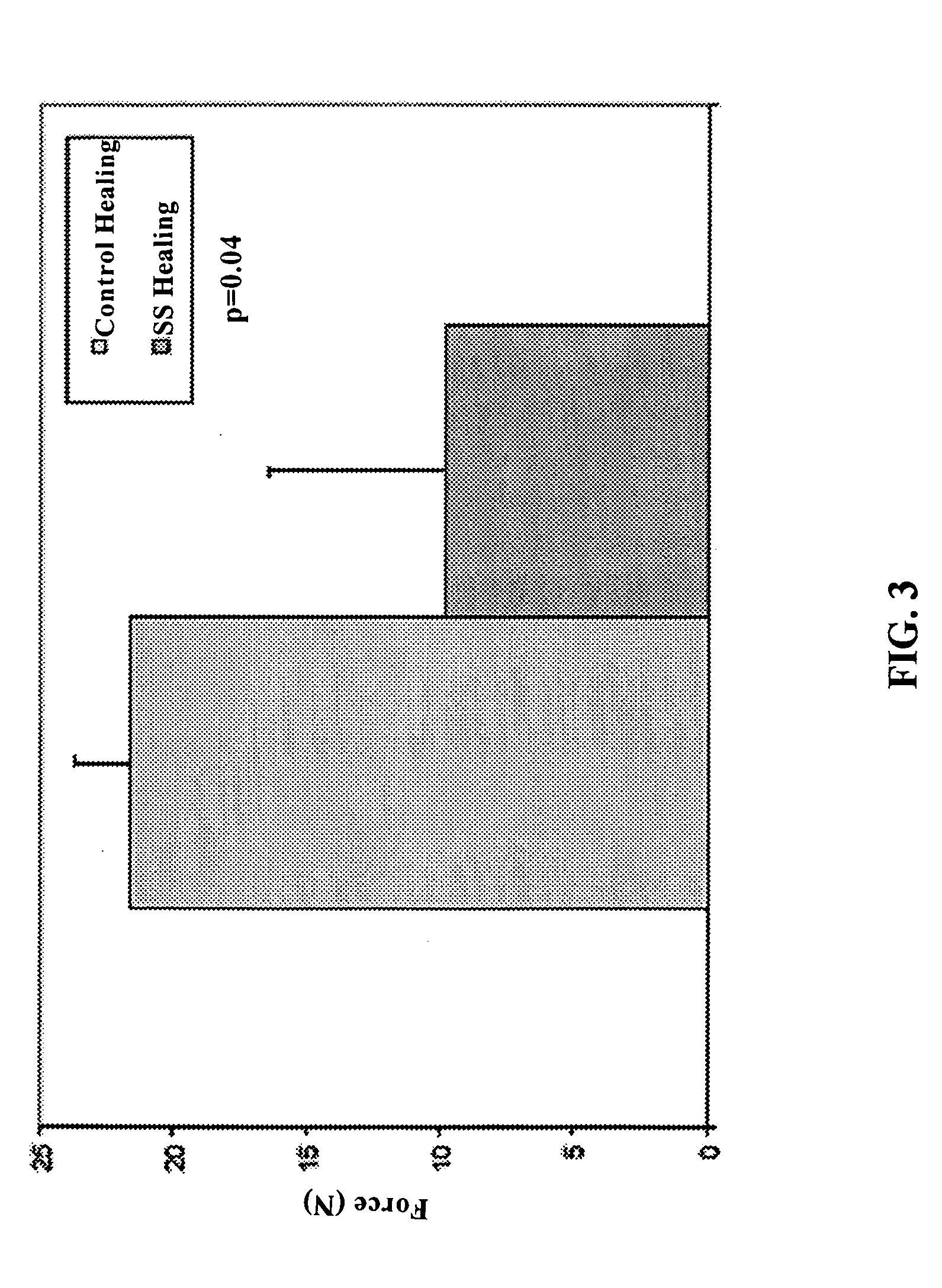
Surgical Sympathectomy Impairs Healing in a Rat MCL Model

[0088] The purpose of this Example was to investigate the role of sympathetic nerves on MCL healing in another rat model.

[0089] Surgical Procedure: Thirteen female Wistar rats (306 g to 339 g) were divided into two groups: surgical sympathectomy (SS) or sham control. All rats were anesthetized with isofluorane as described in Example 1. A transperitoneal approach was used. The lower abdomen was shaved and prepared for surgery. A skin incision approximately 25 mm long was made. Fascia and abdominal muscle were incised. Abdominal organs were displaced. With the aid of a dissecting microscope, fascia surrounding the deep muscle tissue was separated (with minimal disruption to the muscle tissue) to expose the post-ganglionic sympathetic nerves. The L1-4 nerves were isolated and transected (with removal of a 5 mm segment of the nerves) in the surgical sympathectomy group only. In both groups, the deep muscle tissue was returned t...

example 3

Local Delivery of Neuropeptides

[0097] Local delivery of NPs and NP inhibitors is important to identify the specific roles of individual NPs on healing tissues. An animal model for the local delivery of NPs has been developed and is presented in this Example. Briefly, NP infused mini-osmotic pumps were implanted subcutaneously into the back of rats. A small catheter running from the pump to an intramuscular space above the MCL was secured to the rat.

[0098] To test whether sensory NPs improve healing in femoral nerve transection (FNT) ruptured MCLs, three female Wistar rats (199 g to 203 g) were given SP locally to the MCL. Substance P (103 pg / μl) was infused into a miniosmotic pump and was delivered to a ruptured MCL of a FNT rat (see surgical procedure in Example 1 above). Within minutes of surgical intervention, all rats recovered and had normal movement and behavior (grooming, feeding, etc.). Mechanical testing showed SP-supplemented FNT MCLs had a much greater failure force (74...


PUM

PropertyMeasurementUnit
strengthaaaaaaaaaa
cartilage strengthaaaaaaaaaa
mechanicalaaaaaaaaaa

Abstract

Disclosed are a method and a corresponding pharmaceutical composition for treating damaged cartilage and subchondral bone. Neurogenic compounds in general and neuropeptides in particular have been found to be highly effective in stimulated repair of cartilage and bone damaged due to traumatic injury, ligament disease, and disuse. Preferred active ingredients for use in the method and corresponding pharmaceutical composition include calcitonin gene-related peptide (CGRP), cholecystokinin (CCK), dynorphin, enkephalin, galanin, neuropeptide Y (NPY), neurotensin, somatostatin, substance P (SP), thyrotropin-releasing hormone (TRH), vasoactive intestinal peptide (VIP).

Description

CROSS-REFERENCE TO RELATED APPLICATION [0001] This application is a continuation-in-part of co-pending application Ser. No. 11 / 063,627, filed Feb. 23, 2005, which claims priority to provisional application Ser. No. 60 / 547,193, filed Feb. 24, 2004; this application also claims priority to provisional application Ser. No. 60 / 679,560, filed May 10, 2005. The contents of all three of these prior applications are incorporated herein.FEDERAL FUNDING [0002] This invention was made with United States government support awarded by the following agency: NSF 9907977. The United States has certain rights in this invention.REFERENCES AND INCORPORATION BY REFERENCE [0003] Complete bibliographic citations for the references cited herein are contained in a section titled “References,” immediately preceding the claims. All of the documents listed in the “References” section and in the text are incorporated herein. FIELD OF THE INVENTION [0004] The invention is generally directed to a method of treat...

Claims


Application Information

Patent Timeline
Patent Type & Authority Applications(United States)
IPC IPC(8): A61F2/28A61K38/31A61K38/22
CPCA61K38/046A61K38/066A61K38/2207A61K38/225A61K38/31A61L27/3843A61L27/3608A61L27/3612A61L27/3641A61L27/3645A61L27/3683A61L27/227A61L2430/06Y10S530/839Y10S530/84
Inventor VANDERBY, RAYGRORUD, KELLEY W.
Owner WISCONSIN ALUMNI RES FOUND
Learn More