
Method of testing a medical imaging device

a portable xray and imaging device technology, applied in the field of portable xray devices, can solve problems such as system damage, system damage, and system damage, and achieve the effect of avoiding system damage, avoiding system damage, and avoiding system damag

Active Publication Date: 2006-10-26
GENERAL ELECTRIC CO
View PDF12 Cites 16 Cited by
  • Summary
  • Abstract
  • Description
  • Claims
  • Application Information

AI Technical Summary

Benefits of technology

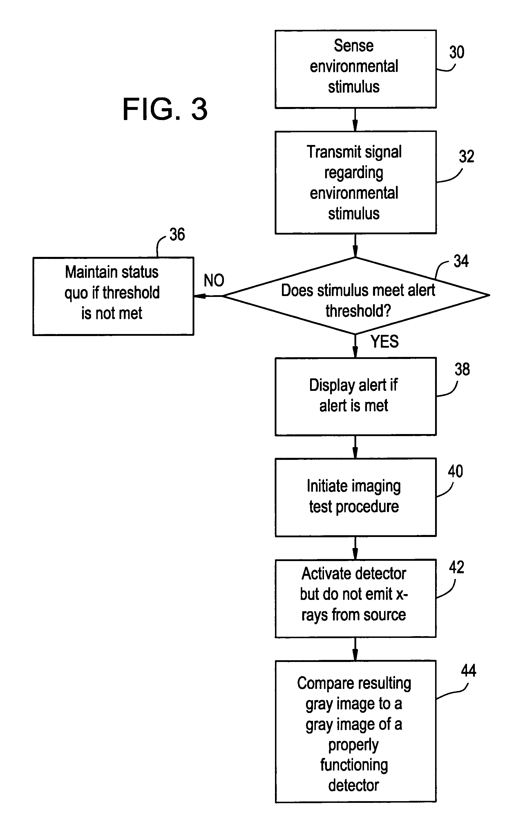
[0006] Certain embodiments of the present invention provide a method of testing a portable x-ray device that includes sensing an environmental stimulus experienced by the portable x-ray device, transmitting a signal related to the environmental stimulus to a processing unit, determining whether the signal meets an alert threshold, activating a detector of the portable x-ray device if the signal meets the alert threshold, producing a gray image through the

Problems solved by technology

The imaging system is susceptible to damage because it is portable and easily moved to different locations.
For example, the system may be tipped over, dropped and / or struck, thereby causing damage to the detector or other components.
For example, if the resulting image contains various types of distortion, the operator determines that the system has been damaged and needs repair.
Testing the system through a full imaging process, however, takes time, and exposes the environment to x-rays.

Method used


Image

  • Method of testing a medical imaging device
  • Method of testing a medical imaging device
  • Method of testing a medical imaging device

Examples


Embodiment Construction

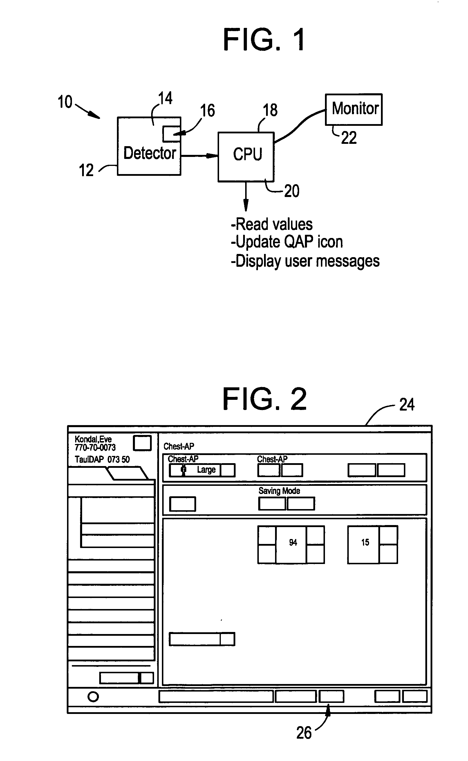
[0013]FIG. 1 illustrates a simplified block diagram of a portable x-ray imaging system 10, according to an embodiment of the present invention. The x-ray system 10 includes a portable x-ray device 12 including a detector 14 having a sensor 16 mounted or otherwise secured thereto. The detector 14 and the sensor 16 are in communication with a computer 18 having a central processing unit (CPU) 20, which is also in communication with a monitor 22. The components of the system 10 may be in communication with each other through wired or wireless connections.

[0014] The sensor 16 may be any type of sensing device that is configured to detect movement. For example, the sensor 16 may be an accelerometer. If the x-ray device 12 is tipped over, dropped or struck, the sensor 16 measures the physical shock, jolt, etc. absorbed by the x-ray device 12. The CPU 20 then receives a signal from the sensor 16 related to the measured shock. The CPU 20 is programmed to determine whether a threshold alert...


PUM


Abstract

A method of testing a portable x-ray device includes sensing an environmental stimulus experienced by the portable x-ray device, transmitting a signal related to the environmental stimulus to a processing unit, determining whether the signal meets an alert threshold, activating a detector of the portable x-ray device if the signal meets the alert threshold, producing a gray image through the activating step, comparing the gray image produced through the activating step with a control gray image corresponding to a properly functioning detector.

Description

RELATED APPLICATIONS [0001] This application relates to and claims priority benefits from U.S. Provisional Patent Application No. 60 / 673,897 entitled “Method Of Testing A Medical Imaging Device,” filed Apr. 22, 2005, which is incorporated by reference herein in its entirety.BACKGROUND OF THE INVENTION [0002] Embodiments of the present invention provide a method of assuring proper operation of a medical imaging device, and, more particularly, to a method of testing a portable x-ray device to determine if it is operating properly. [0003] Portable x-ray systems are used to image anatomical structures within various settings. For example, a technician may use a portable x-ray system to image an object of interest, such as a forearm, in a hospital bedroom setting. The technician may image the object with the x-ray system, and then leave the room with the portable system to image another patient in another location. The imaging system is susceptible to damage because it is portable and ea...

Claims


Application Information

Patent Timeline
IPC IPC(8): G01D18/00
CPCA61B6/583
Inventor LIU, JAMES ZHENGSHEKUKRER, OLGUNKUMP, KENNETH SCOTTOMERNICK, JON C.
Owner GENERAL ELECTRIC CO
Learn More