
Engineering vascularized muscle tissue

a technology of vascularized muscle tissue and engineered constructs, which is applied in the direction of artificial cell constructs, skeletal/connective tissue cells, biocide, etc., can solve the problems of complex tissue engineering, inability to vascularize the tissue in vitro, and inability to achieve thick, highly vascularized tissues

Inactive Publication Date: 2006-09-07
MASSACHUSETTS INST OF TECH
View PDF4 Cites 36 Cited by
  • Summary
  • Abstract
  • Description
  • Claims
  • Application Information

AI Technical Summary

Benefits of technology

[0021] In another aspect, the invention is a method of producing a tissue engineered construct. The method includes providing a population of endothelial cells, providing a population of muscle cells, seeding the endothelial cells and the muscle cells on a three-dimensional support matrix, and culturing the seeded cell support matrix in a predetermined medium for a predetermined period of time. Seeding may include suspending the muscle cells and the endothelial cells in growth-factor reduced Matrigel and absorbing a predetermined amount of the suspen

Problems solved by technology

One of the major obstacles in engineering thick, complex tissues such as muscle is the need to vascularize the tissue in vitro.
Although this approach has been useful in many tissues, it has not been as successful in thick, highly vascularized tissues such as the muscle (Saxena, et al., Tissue Eng (1999) 5, 525-532; Neumann, et al., Microvasc Res (2003) 66, 59-67; Bach, et al., Clin Plast Surg (2003) 30, 589-599).
Biodegradable polymers are not necessarily hydrolytically degradable and may require enzymatic action to fully degrade.

Method used


Image

  • Engineering vascularized muscle tissue
  • Engineering vascularized muscle tissue
  • Engineering vascularized muscle tissue

Examples


examples

Cell Culture

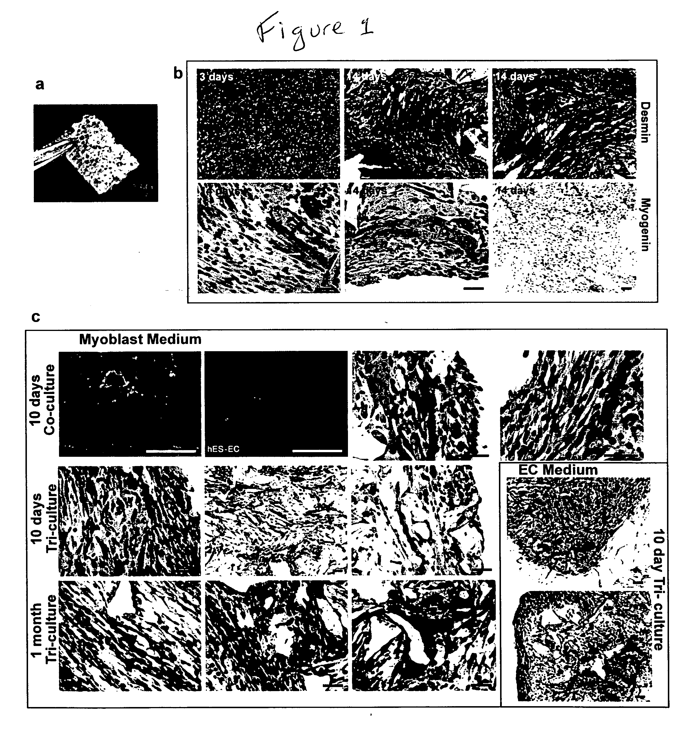
[0052] Mouse myoblast cells (C2C12) (Yaffe, et al., Nature (1977) 270, 725-727; Blau, et al., Science (1985) 230, 758-766, the contents of both of which are incorporated herein by reference) were cultured in DMEM supplemented with 10% fetal bovine serum (FBS), 10% calf serum, and 2.5% HEPES buffer (myoblast medium). Human umbilical vein endothelial cells (HUVEC) were cultured in endothelial cell medium (EGM-2; Cambrex Biosciences). Mouse embryonic fibroblasts (MEF) (Cell Essentials, Boston) were cultured in DMEM supplemented with 10% FBS (embryonic fibroblast medium). HESC-derived CD31+endothelial cells were isolated as described (Levenberg, et al., 2002) and cultured in endothelial cell medium.

Polymer Scaffolds

[0053] Porous sponges composed of 50% poly-(L-lactic acid) (PLLA) and 50% polylactic-glycolic acid (PLGA) were fabricated as described (Levenberg, et al., 2003) with pore sizes of 225-500 μm and 93% porosity. The PLGA was selected to degrade quickly (˜3 weeks...


PUM


Abstract

A tissue engineered construct. The construct includes endothelial cells, muscle cells, and a three-dimensional support matrix on which the endothelial cells and the myoblasts are seeded.

Description

[0001] This application claims priority from U.S. Patent Applications Nos. 60 / 650,427, filed Feb. 4, 2005, and 60 / 691,609, filed Jun. 17, 2005, the entire contents of both of which are incorporated herein by reference[0002] This work was supported by NIH grants HL60435 and EY05318. The government may have certain rights in this invention.FIELD OF THE INVENTION [0003] This invention relates to vascularized tissue engineered constructs and methods of making same. BACKGROUND OF THE INVENTION [0004] One of the major obstacles in engineering thick, complex tissues such as muscle is the need to vascularize the tissue in vitro. Vascularization in vitro could maintain cell viability during tissue growth, induce structural organization and promote vascularization upon implantation. Many past approaches to engineering new tissue have relied on the host for vascularization. Although this approach has been useful in many tissues, it has not been as successful in thick, highly vascularized tissu...

Claims


Application Information

Patent Timeline
IPC IPC(8): A61K35/12C12N5/06C12N5/08C12N5/071C12N5/077
CPCC12N5/0657C12N5/0658C12N5/0697C12N2502/28
Inventor LEVENBERG, SHULAMITROUWKEMA, JEROENLANGER, ROBERT S.
Owner MASSACHUSETTS INST OF TECH
Learn More