
Tissue sensor and uses thereof

a technology of tissue sensor and electrode, applied in the field of tissue sensor, to achieve the effect of efficient and accura

Inactive Publication Date: 2006-05-18
MYOMICS
View PDF7 Cites 79 Cited by
  • Summary
  • Abstract
  • Description
  • Claims
  • Application Information

AI Technical Summary

Benefits of technology

[0078] Combining the data obtained through the sensor with gene expression data can provide powerful insights into the activity of known or new drug agents on such tissues. Gene activity can be monitored by, for example, PCR targeting one or a number of genes, known or unknown. In one aspect, nucleic acid derived from such tissues can be used to probe a microarray, thereby providing a genetic expression profile for that time point. Other approaches to genetic profiling, e.g., approaches based on differential display or similar methods are known in the art. By obtaining simultaneous genetic expression data, the pathways influenced by a given drug or stimulus that affects mechanical function can also be identified. The data obtained by monitoring a number of similar or identical tissues for a parameter such as muscle contraction, relaxation, etc., over time using a tissue / sensor combination as described herein can also be combined with data regarding protein expression profile or proteomic analysis in a similar fashion.
[0079] In part because of the number of tissues that can be simultaneously or at least contemporaneously monitored, as well as because tissues described herein can be maintained for extended periods of time, the tissue / sensor combinations described herein are well suited for long term studies, e.g., on the order of days, weeks, or even months. As such, they can provide data regarding tissue function in response to a drug or stimulus and ways in which the response or the tissue can change with long-term or repeat exposure to the drug or stimulus. This can be predictive of, for example, the long term effects of a drug or stimulus on the organization or function of a tissue. Such long term studies can also identify, for example, activities of known drugs on tissues which may not become apparent in shorter term studies. Thus, the tissue-sensor combinations described herein can permit the identification of new beneficial uses of known drugs, e.g., where a drug or compound known to be tolerated in vivo is found to have an activity not previously appreciated. Such long term studies can also potentially identify previously unappreciated harmful effects of known or new drugs. This long-term predictive aspect becomes even more powerful when coupled with the ability to monitor the genetic or proteomic profile of tissues from the same experiment at times corresponding to the mechanical or physical measurements provided by a sensor.
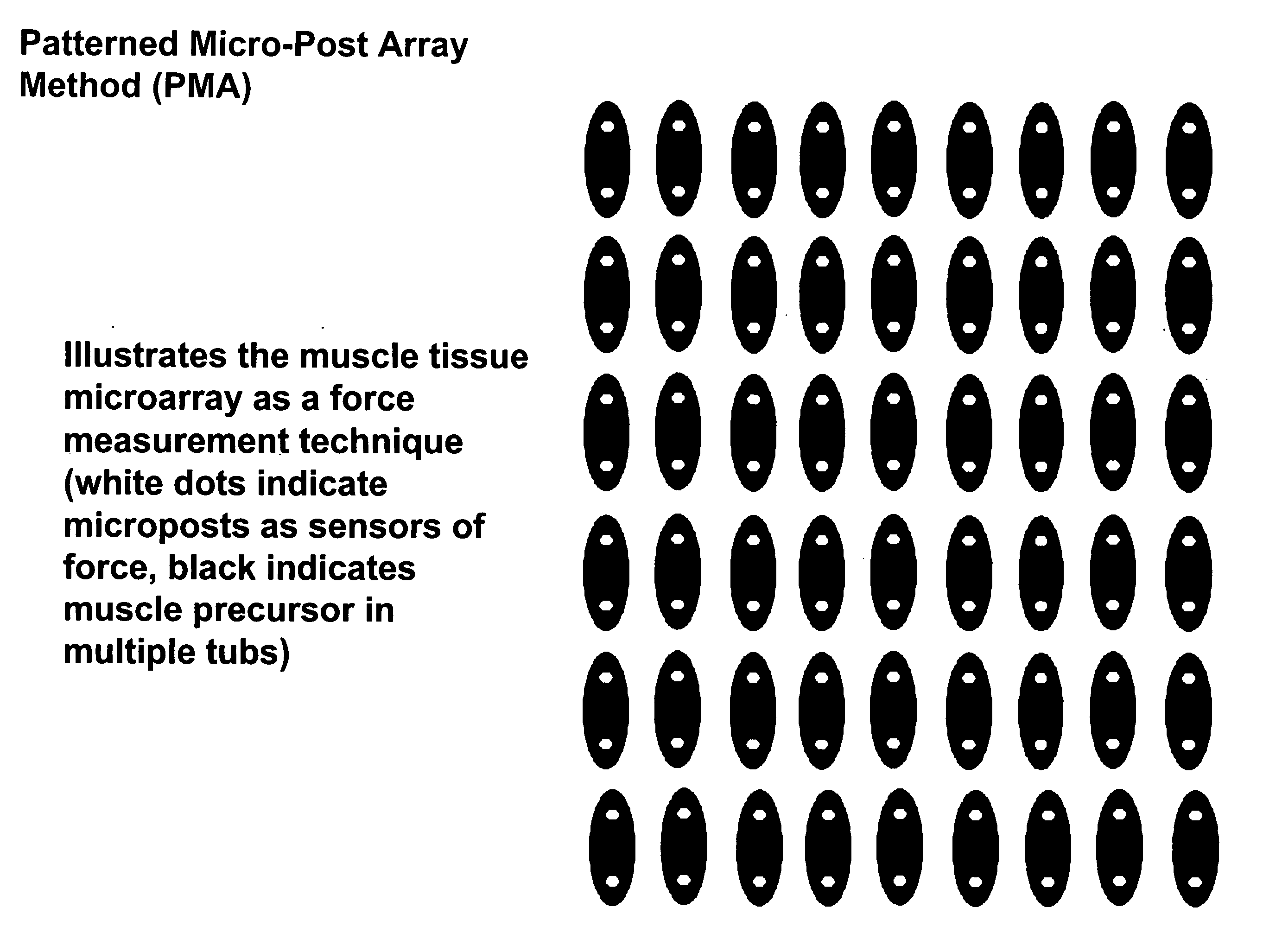
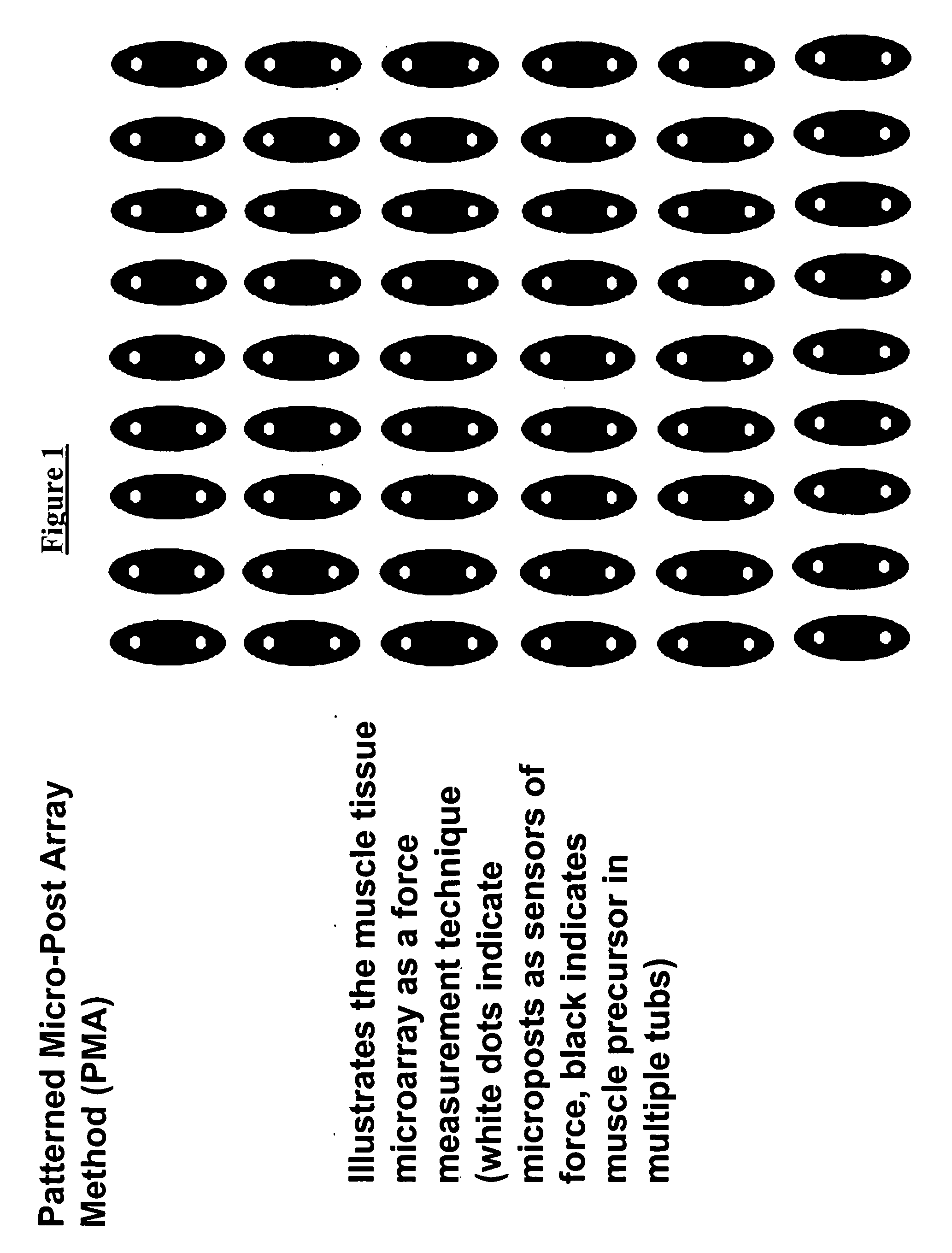
[0081] In a preferred embodiment of the kit the container comprises a culture plate containing a plurality of tubs, wherein each tub contains a tissue or a plurality of tissues in medium and under conditions wherein the tissue is viable, long-term. The tubs can be isolated from other tubs, as, for example, separate wells in a multi-well culture plate, or, alternatively, a plurality of tubs can be present in a single well. In either instance, the tubs can be arranged in an array, thereby facilitating more rapid gathering of information regarding the effect(s) of a compound or compounds.
[0083] Further features and advantages of the compositions and methods described herein include the following. The organized tissue aspect described herein provides a more in vivo-like culture system for screening the activity of biological compounds and offers advantages over disorganized tissue. For example, poorly differentiated cells respond differently to compounds as compared to organized cells in vivo. Also provided are methods for screening a bioactive compound in a tissue which reflects the in vivo cellular organization and gross morphology of the natural in vivo tissue. This organized tissue system offers an efficient and accurate method for screening candidate bioactive compounds for desired biological effects in vitro and in vivo, and permits screening on a long-term, rather than a short-term basis.

Problems solved by technology

In addition, certain cell types (e.g. muscle, fibroblasts, bone and cartilage) are anchorage dependent, and when these adherent cells grown as a monolayer are spontaneously released into the culture medium, they will die.

Method used


Image

  • Tissue sensor and uses thereof
  • Tissue sensor and uses thereof
  • Tissue sensor and uses thereof

Examples


example 1

Preparation of a Tissue in Combination with a Sensor

[0265] A tissue in combination with a sensor can be prepared as follows, which exemplifies a preparation using muscle tissue.

[0266] To produce skeletal muscle organoids, primary avian, rat or human muscle stem cells or immortalized murine muscle cells, were suspended in a solution of collagen and Matrigel™ which was maintained at 4° C. to prevent gelling. The cell suspension was then placed in a vessel with tissue attachment surfaces coupled to an interior surface at each end of the vessel. The vessel was positioned in the bottom of a standard cell culture chamber. Following two to four hours of incubation at 37° C., the gelled cell suspension was covered with fresh culture medium (renewed at 24 to 72 hour intervals) and the chamber containing the suspended cells was maintained in a humidified 5% CO2 incubator at 37° C. throughout the experiment.

[0267] Between the second and sixth day of culture, the cells were found to be organ...

example 2

Use of a Tissue Sensor for Screening a Compound for Bioactivity

[0278] A tissue and sensor, e.g., a muscle tissue and sensor prepared as described herein can be used to screen for bioactive compounds, for example, as follows.

[0279] An array of tissues formed in isolated wells in, e.g., a 384 or 96 well tissue culture dish is contacted with test compound by adding the test compound(s) to the wells, either individually or with a multipipettor. Readings from the sensor before and after (e.g., 1 second, 5 seconds, 30 seconds, 1 min, 5 min, 1 hr, etc. after) addition of the test compound determine the bioactivity of the compound. For example, contraction or relaxation of muscle tissue occurring after introduction of the compound is indicative that the compound is bioactive on the tissue.

[0280] The method of performing the assay will vary depending upon the particular assembly of the sensor and tissue. For example, where the tissue is independent from the sensor, a single sensor can be ...

example 3

Use of a Tissue Sensor for Screening a Library of Compounds for Bioactivity

[0281] A library of compounds is screened by, for example, preparing an array of tissues in combination with a sensor, and then contacting different members of the array with different members of the library of compounds. This can be achieved, for example, where a library of compounds is added to different wells of an array of wells comprising tissue. The tissue can be in combination with a sensor within the well, or a sensor or set of sensors can be moved from well to well, depending upon the type of sensor used.

[0282] In one aspect, an array of bubble-type sensor-muscle tissue assemblies is immersed in wells of a plate comprising members of the library. Pressure readings from the array of sensor-tissue assemblies provides a read out on the bioactivity of members of the library. In one embodiment, which is applicable to any of the library screening approaches described herein, the library is split up into ...


PUM

PropertyMeasurementUnit
molecular massaaaaaaaaaa
lengthaaaaaaaaaa

Abstract

Described are assemblies for screening a compound for bioactivity, the assemblies comprising a tissue and a sensor. A change in a biological parameter is measured by the sensor, such that a change in a parameter occurring when the tissue is contacted with a candidate compound is detected by the sensor. Assemblies provided herein include single sensor / tissue assemblies and arrays of such assemblies, including plates comprising tissues in combination with one or more sensors. Also provided are methods of screening a compound using tissue / sensor tissue assemblies as described.

Description

[0001] This application is a Continuation In Part of U.S. patent application Ser. No. 10 / 241,618, filed Sep. 11, 2002, which is a Continuation of U.S. patent application Ser. No. 09 / 252,324, filed Feb. 18, 1999, now abandoned, which claims the priority of U.S. Provisional application No. 60 / 075,054, filed Feb. 18, 1998 and U.S. Provisional application No. 60 / 086,370, filed May 22, 1998.FIELD OF THE INVENTION [0002] The invention relates to the measurement of a parameter of a tissue and to measurement of bioactivity of a compound on such a tissue. BACKGROUND OF THE INVENTION [0003] In vitro screening of compounds for biological activity has been disclosed in the prior art as assays, for example, in which monolayers of tissue cultured cells are exposed to a candidate compound and a biological response in the cells is measured. For example, monolayers of disorganized muscle fibers have been shown to respond to anabolic growth factors. See Vandenburgh et al. (Vandenburgh et al., Am. J. ...

Claims


Application Information

Patent Timeline
Patent Type & Authority Applications(United States)
IPC IPC(8): C12Q1/68G01N1/30C12M1/34
CPCB01L3/5085B01L3/5088B01L2300/0636B01L2300/0819G01N33/5088G01N33/6887
Inventor BENESCH, FRANKBARBATA, VICTORIA MARGITVALENTINI, ROBERT FRANCISVANDENBURGH, HERMAN H.CRAWFORD, GREGORY PHILIP
Owner MYOMICS
Learn More