Protein tyrosine kinase substrate LAT and its use in the indentification of (ANT)agonists of the kinase
a kinase substrate and protein tyrosine technology, applied in the field of identifying and testing tyrosine kinase signaling pathway agonists and antagonists, can solve the problems of limited clinical indications of cyclosporin (and the more recently developed fk506), and achieve enhanced signaling, prevent association, and prevent tyrosine phosphorylation
- Summary
- Abstract
- Description
- Claims
- Application Information
AI Technical Summary
Benefits of technology
Problems solved by technology
Method used
Image


Examples
example 1
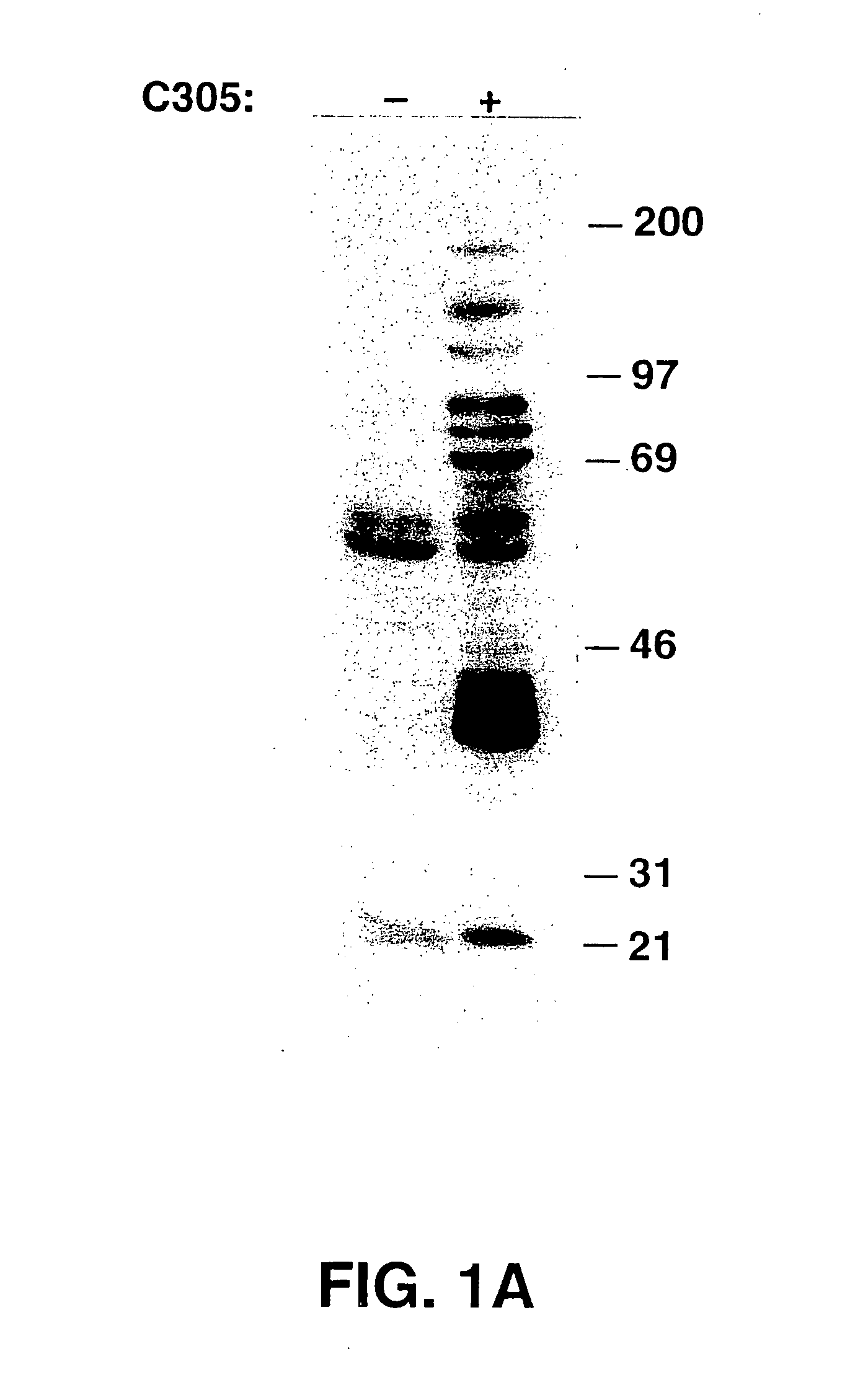
[0265] In this example, the purification process of LAT is described. Stimulation of the TCR on the Jurkat human T cell line by crosslinking with either C305 (anti-TCRβ, See FIG. 1A) or OKT3 (anti-CD3ε, not shown) monoclonal antibodies induced the tyrosine phosphorylation of multiple proteins, most prominently, p36-38. To define the role of p36-38 in T cell signaling, the protein was purified from activated Jurkat cells. Since, p36-38 appears to be membrane-associated, membrane fractions were prepared from OKT3 stimulated Jurkat cells and extracted with Brij97 detergent. The extracted membrane proteins were then heat-treated, which induced precipitation of about ⅔ of the protein, but not of p36-38. Anti-phosphotyrosine antibodies were used for affinity purification of phosphotyrosine-containing proteins. These were then specifically eluted with phenyl phosphate, concentrated, and resolved on SDS-PAGE. The p36-38 band was excised, digested in gel with trypsin, and the resultant trypt...
example 2
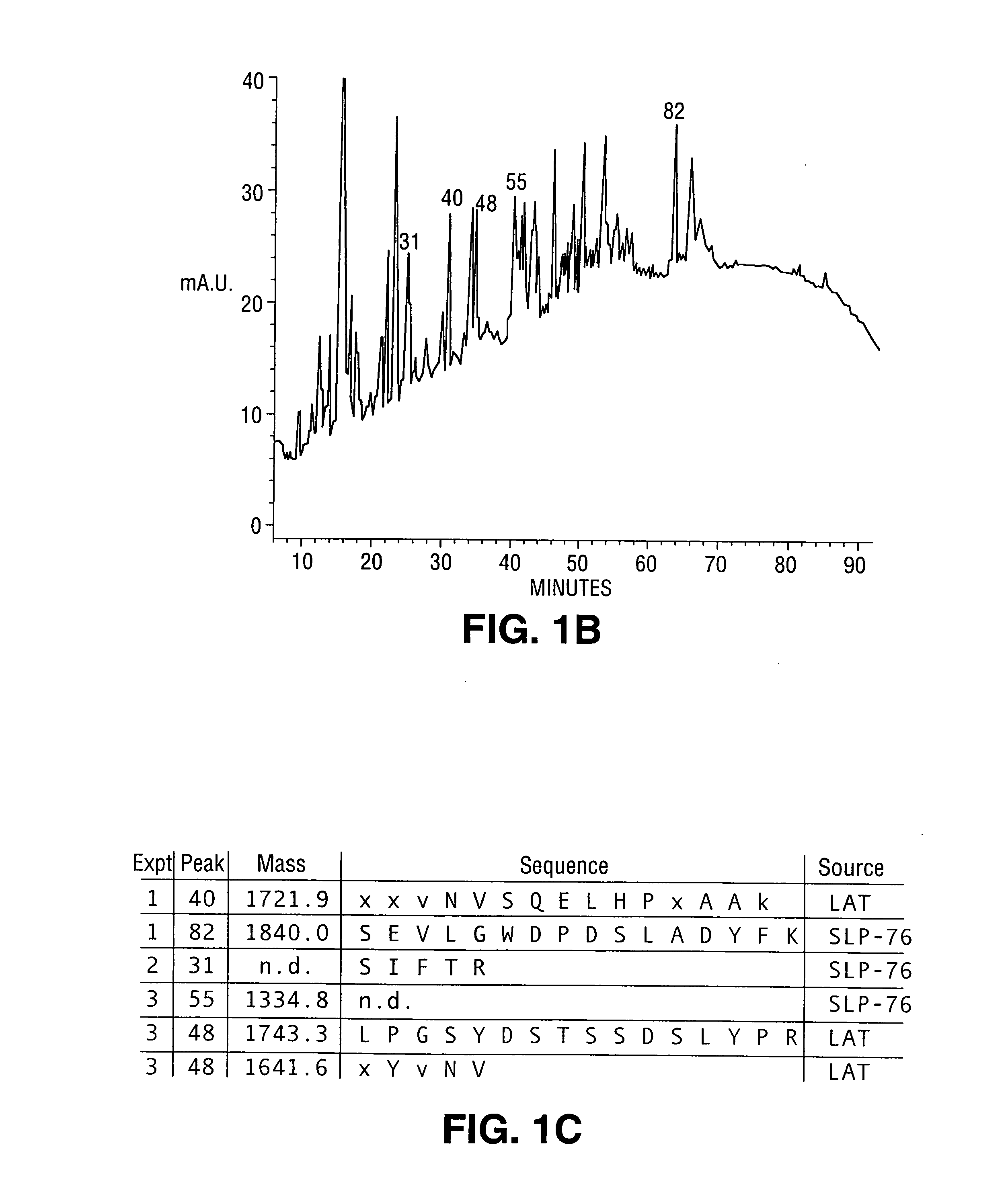
[0266] In this example, peptide sequencing and cDNA cloning of the LAT protein is described. Peptides from five HPLC fractions were either microsequenced or subjected to mass spectrometry. The peptide from fraction 40 had a molecular weight of 1721.9 daltons (See FIG. 1C). The residues at position 1, 2, 3, 11 and 15 could not be assigned unambiguously. The available sequence was then used to search the Genbank data base with the BLAST algorithm, and an EST clone from human fetal heart was found to encode the peptide fragment. Review of the Edman sequence data at the ambiguous positions was consistent with the EST sequence. The predicted molecular weight of the putative tryptic fragment predicted from the EST sequence was 1641.8 daltons. The 80.1 daltons difference suggested that a phosphate moiety of this molecular weight could be present at the Tyr residue. Two peptides from peaks 82 and 31 were shown by sequence analysis to be derived from the known tyrosine kinase substrate SLP-7...
example 3
[0272] In this example, tissue and cellular distribution of the LAT protein is described. Northern blot analysis was performed with poly (A)+ RNAs isolated from different adult human tissues. Two transcripts (2.0 and 1.6 Kb) were seen predominantly in thymus, peripheral blood, and at a low level of expression in spleen. There were no transcripts of LAT found in other tissues (See FIG. 3A and data not shown). LAT expression was assayed in human and rat cell lines of hematopoietic origin. LAT mRNA was only found in Jurkat, YT (NK-like cells) and RBL (a rat mast cell line) (See FIG. 3B). From these data, it was concluded that LAT is expressed in T cells, NK cells, and mast cells, but not in B cells (Raji and Jiyoye) or monocytes (THP1).
[0273] The cellular localization of LAT was analyzed by immunofluorescence in COS cells, which were transiently transfected with LAT cDNA, fixed, permeabilized and incubated sequentially with anti-LAT and TRITC-conjugated goat anti-rabbit antibodies. Vi...
PUM
| Property | Measurement | Unit |
|---|---|---|
| Tm | aaaaa | aaaaa |
| temperature | aaaaa | aaaaa |
| temperature | aaaaa | aaaaa |
Abstract
Description
Claims
Application Information
 - R&D
- Intellectual Property
- Life Sciences
- Materials
- Tech Scout
- Unparalleled Data Quality
- Higher Quality Content
- 60% Fewer Hallucinations
Browse by: Latest US Patents, China's latest patents, Technical Efficacy Thesaurus, Application Domain, Technology Topic, Popular Technical Reports.
© 2025 PatSnap. All rights reserved.Legal|Privacy policy|Modern Slavery Act Transparency Statement|Sitemap|About US| Contact US: help@patsnap.com