
Device and method for monitoring body fluid and electrolyte disorders

Inactive Publication Date: 2006-01-26
TYCO HEALTHCARE GRP LP
View PDF88 Cites 232 Cited by
  • Summary
  • Abstract
  • Description
  • Claims
  • Application Information

AI Technical Summary

Benefits of technology

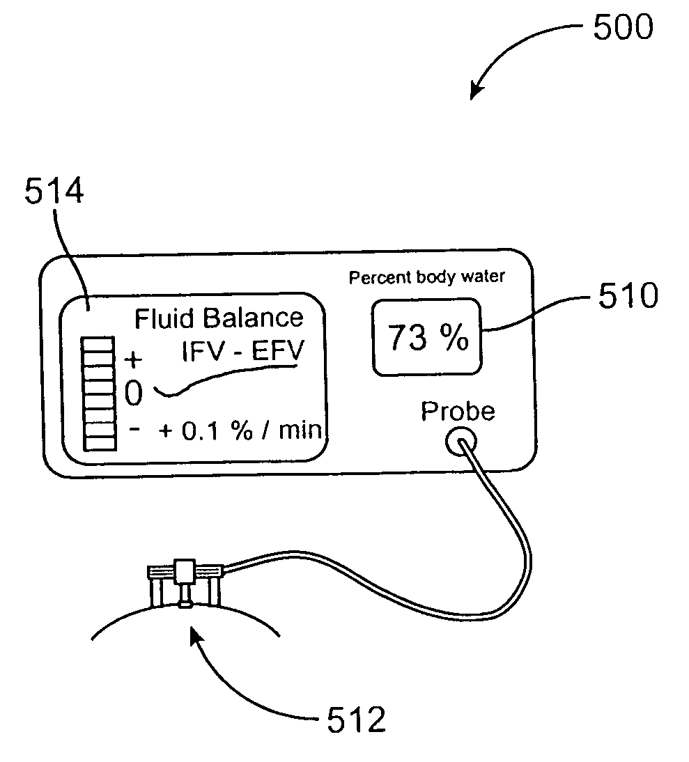
[0006] The difference between the fraction of water in the intravascular fluid volume (“IFV”) and extravascular fluid volume (“EFV”) compartments are also determined using a differential method that takes advantage of the observation that pulsations caused by expansion of blood vessels in the skin, as the heart beats, produce changes in the received radiation at a particular wavelength that are proportional to the difference between the effective absorption of light in the blood and the surrounding tissue. This difference, integrated over time, provides a measure of the quantity of the fluid that shifts into and out of the capillaries. A mechanism for mechanically inducing a pulse is built into the device to improve the reliability of measurements of IFV−EFV under weak-pulse conditions.

Problems solved by technology

The maintenance of body fluid balance is of foremost concern in the care and treatment of critically ill patients, yet physicians have access to few diagnostic tools to assist them in this vital task.
Dehydration of infants and children suffering from diarrhea can be life-threatening if not recognized and treated promptly.
None of these variables alone, however, is a direct and quantitative measure of water retention or loss.
It is, however, an invasive technique that requires blood sampling.
The rather poor accuracy of the technique seen in many studies point to unresolved deficiencies of these designs when applied in a clinical setting.

Method used


Image

  • Device and method for monitoring body fluid and electrolyte disorders
  • Device and method for monitoring body fluid and electrolyte disorders
  • Device and method for monitoring body fluid and electrolyte disorders

Examples


Embodiment Construction

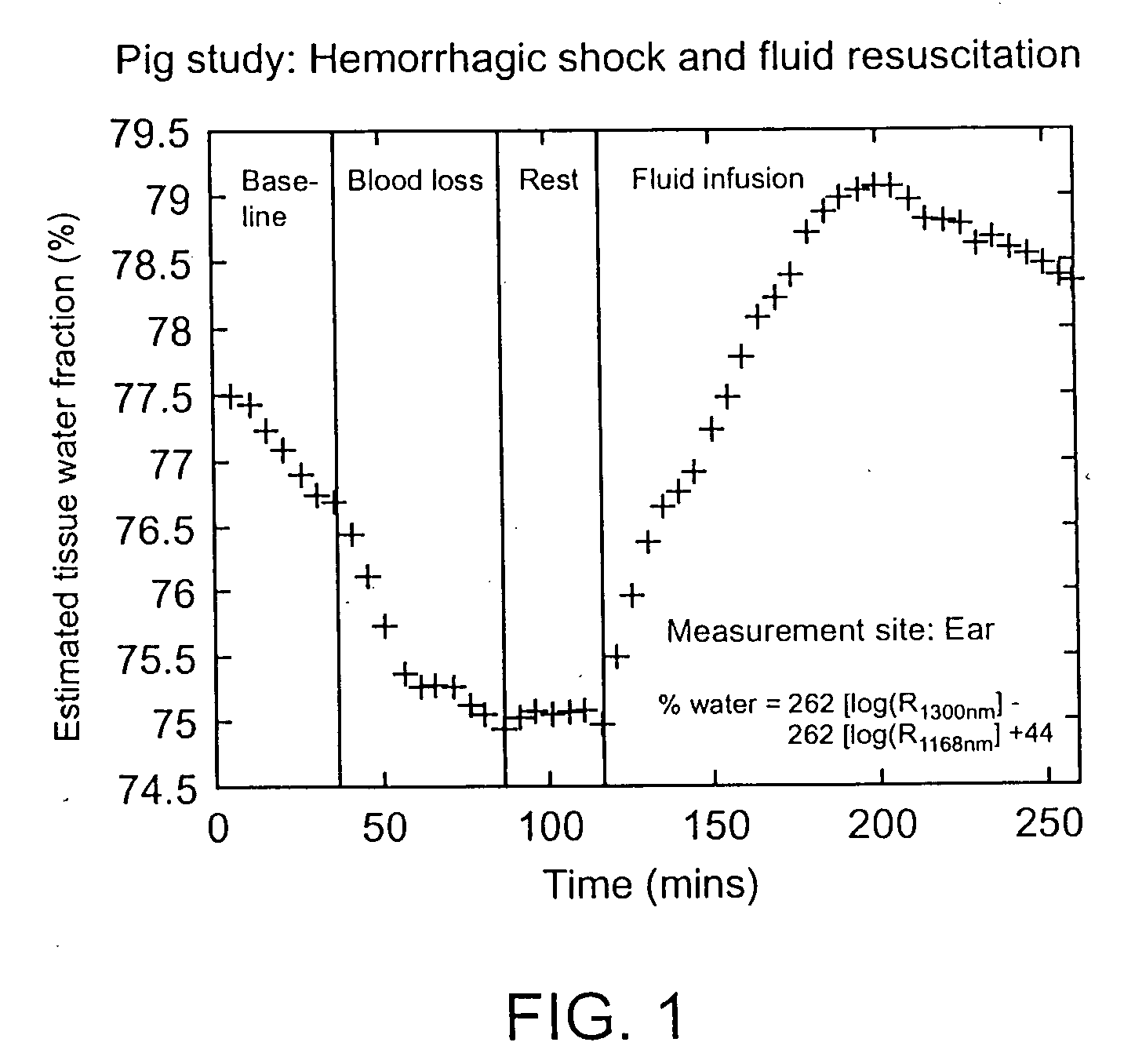
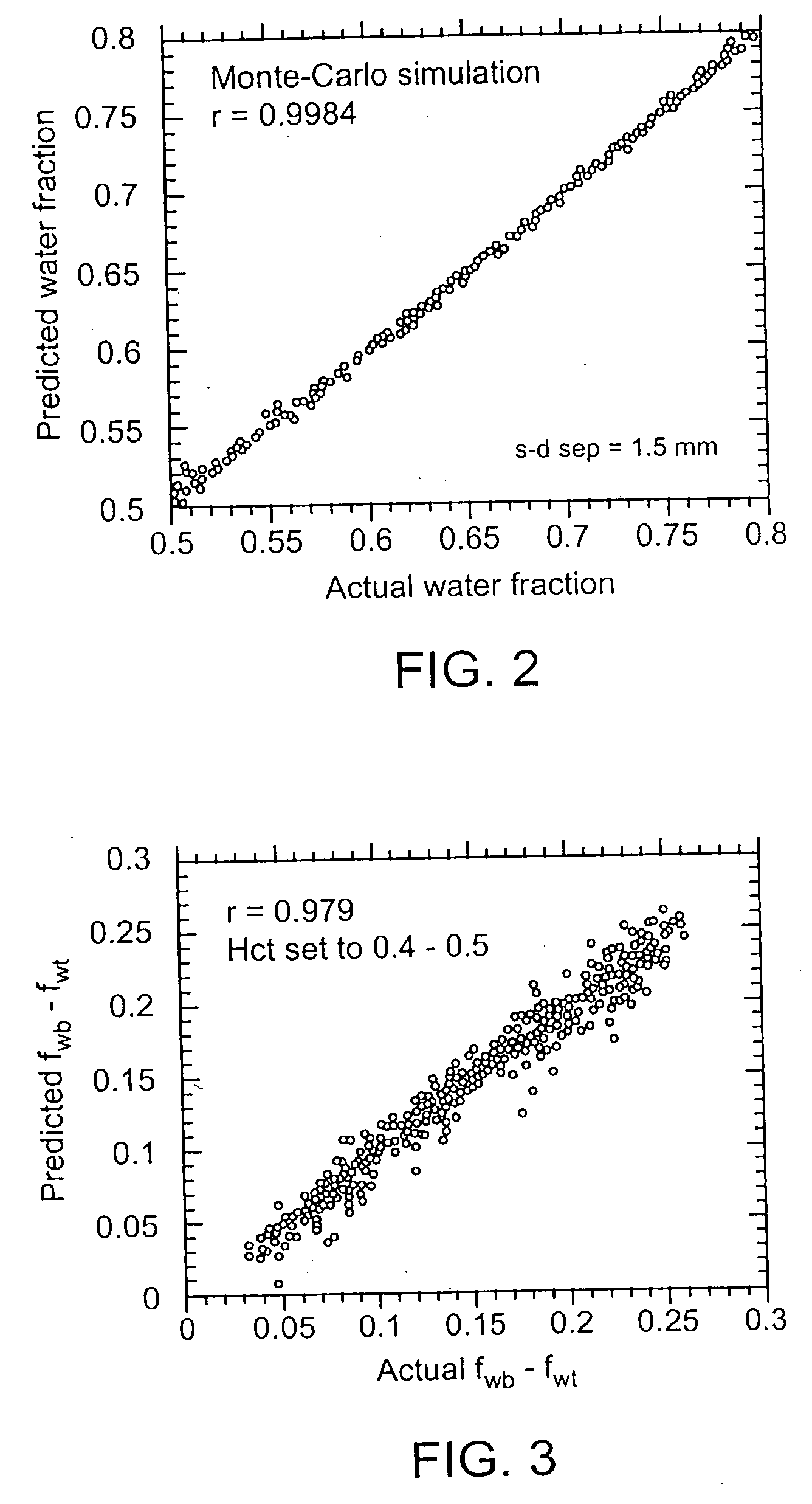
[0014] Embodiments of the present invention overcome the problems of invasiveness, subjectivity, and inaccuracy from which previous methods for body fluid assessment have suffered. The method of diffuse reflectance near-infrared (“NIR”) spectroscopy is employed to measure the absolute fraction of water in skin. An increase or decrease in the free (non protein-bound) water content of the skin produces unique alterations of its NIR reflectance spectrum in three primary bands of wavelengths-(1100-1350 nm, 1500-1800 nm, and 2000-2300 nm) in which none-heme proteins (primarily collagen and elastin), lipids, and water absorb. According to the results of numerical simulations and experimental studies carried out by the inventor, the tissue water fraction fw, defined spectroscopically as the ratio of the absorbance of water and the sum of the absorbances of none-heme proteins, lipids, and water in the tissue, can be measured accurately in the presence of nonspecific scattering variation, te...


PUM


Abstract

A device and a method for measuring body fluid-related metrics using spectrophotometry to facilitate therapeutic interventions aimed at restoring body fluid balance. The specific body fluid-related metrics include the absolute volume fraction of water in the extravascular and intravascular tissue compartments, as well as the shifts of water between these two compartments. The absolute volume fraction of water is determined using algorithms where received radiation measured at two or more wavelengths are combined to form either a single ratio, a sum of ratios or ratio of ratios of the form log[R(λ1) / R(λ2)] in which the received radiation in the numerator depends primarily on the absorbance of water and the received radiation in the denominator depends primarily on the absorbance of water and the sum of the absorbances of non-heme proteins, lipids and water in tissue. The difference between the fraction of water in the intravascular fluid volume (“IFV”) and extravascular fluid volume (“EFV”) compartments are also determined using a differential method that takes advantage of the observation that pulsations caused by expansion of blood vessels in the skin as the heart beats produce changes in the received radiation at a particular wavelength that are proportional to the difference between the effective absorption of light in the blood and the surrounding tissue. This difference, integrated over time, provides a measure of the quantity of the fluid that shifts into and out of the capillaries. A mechanism for mechanically inducing a pulse is built into the device to improve the reliability of measurements of IFV−EFV under weak-pulse conditions.

Description

BACKGROUND OF THE INVENTION [0001] The maintenance of body fluid balance is of foremost concern in the care and treatment of critically ill patients, yet physicians have access to few diagnostic tools to assist them in this vital task. Patients with congestive heart failure, for example, frequently suffer from chronic systemic edema, which must be controlled within tight limits to ensure adequate tissue perfusion and prevent dangerous electrolyte disturbances. Dehydration of infants and children suffering from diarrhea can be life-threatening if not recognized and treated promptly. [0002] The most common method for judging the severity of edema or dehydration is based on the interpretation of subjective clinical signs (e.g., swelling of limbs, dry mucous membranes), with additional information provided by measurements of the frequency of urination, heart rate, serum urea nitrogen SUN / creatinine ratios, and blood electrolyte levels. None of these variables alone, however, is a direct...

Claims


Application Information

Patent Timeline
IPC IPC(8): A61B5/00A61B5/024A61B5/145A61B5/1455G01N21/35
CPCA61B5/0048A61B5/0059G01N21/359A61B5/14546A61B5/4878A61B5/02438A61B5/0053
Inventor SCHMITT, JOSEPH M.
Owner TYCO HEALTHCARE GRP LP
Learn More