
Method for preparation of water-soluble and dispersed iron oxide nanoparticles and application thereof

a technology of dispersed iron oxide and nanoparticles, which is applied in the field of preparation of water-soluble and dispersed iron oxide nanoparticles, can solve the problems of unstable magnetic field and adverse health effects, and achieve the effect of easy binding of biomolecules and drugs

Inactive Publication Date: 2005-12-08
NAT CHENG KUNG UNIV
View PDF2 Cites 18 Cited by
  • Summary
  • Abstract
  • Description
  • Claims
  • Application Information

AI Technical Summary

Benefits of technology

[0007] To address the drawbacks of prior arts for making water-soluble and dispersed iron oxide nanoparticles and the limitation of magnetic resonance imaging contrast agents currently on the market, the present invention discloses a technology for preparing highly water-soluble iron oxide in aqueous phase process that displays ...

Problems solved by technology

But when the particle is very small, its magnetic field becomes unstable.
Using improper dosage or formulation of Gd3+ contrast agent might produce adverse health effect.

Method used


Image

  • Method for preparation of water-soluble and dispersed iron oxide nanoparticles and application thereof
  • Method for preparation of water-soluble and dispersed iron oxide nanoparticles and application thereof
  • Method for preparation of water-soluble and dispersed iron oxide nanoparticles and application thereof

Examples


example 1

Preparation of Water-Soluble Fe3O4 Nanoparticles

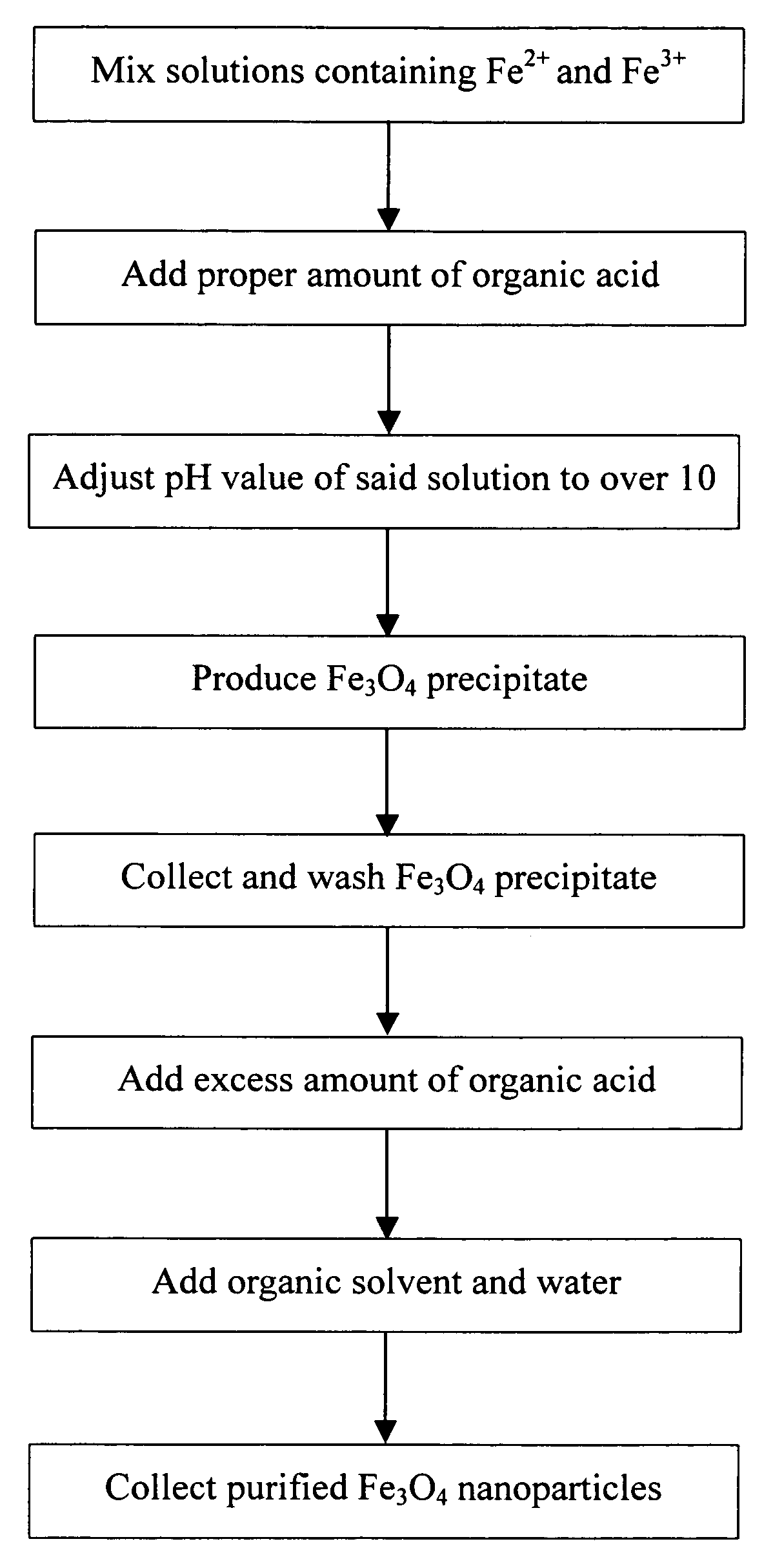
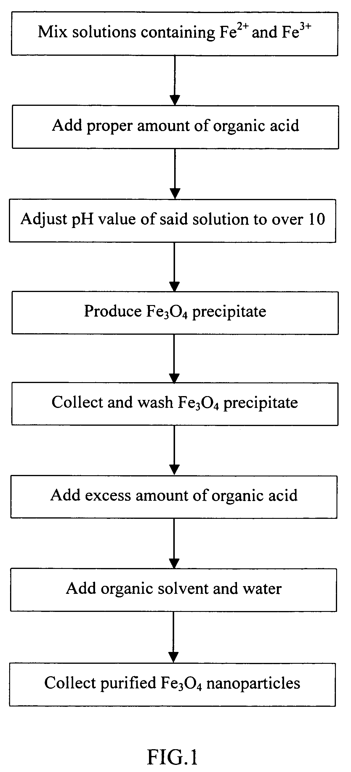
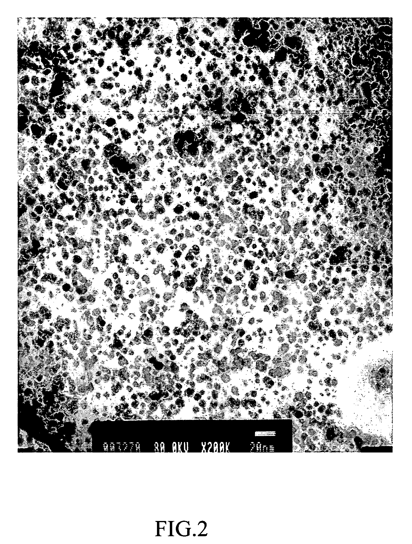
[0026] First mix 0.2 M FeCl2 and 0.1 M FeCl3 in 2 M HCl solution at the volume ratio of 1:4 (FeCl2: FeCl3), then add 1 g of glycine (preferably 0.5˜1.5 g); slowly drip 5 M NaOH solution into the mixture to adjust its pH to over 10 to provide an alkaline environment for Fe3O4 in the solution to precipitate; next agitate for 10 minutes, then wash with D.I. water several times to collect the black precipitate (Fe3O4); next add 3 g of glycine as adherent; agitate 10‥15 minutes and then vibrate for 30 minutes to let the adherent cover the surface of Fe3O4 nanoparticles entirely; subsequently add obtained Fe3O4 nanoparticles to acetone and water mixture to remove excess organic acid adherent; centrifuge at 8000 rpm for 20 minutes to precipitate the Fe3O4 nanoparticles to obtain water-soluble and dispersed Fe3O4 nanoparticles disclosed in the invention. FIG. 2 shows the electron microscope image of resulting Fe3O4 nanoparticles dissolved in ...

example 2

Using Fe3O4 Nanoparticles as MRI Contrast Agent—Injected in Liver

[0027] In this example, Fe3O4 nanoparticles prepared in Example 1 were used as MRI contrast agent. The contrast agent was prepared by dissolving the Fe3O4 nanoparticles in D. I. water, and if necessary, adding to it proper amount of serum or similar body fluid.

[0028]FIG. 3A shows the MRI scan before Fe3O4 nanoparticles were injected into the liver; FIG. 3B shows the MRI scan after the liver was injected with 0.86 μM Fe3O4 nanoparticles. By comparing where the arrows are pointed at in FIGS. 3A and 3B, it is clearly shown that Fe3O4 nanoparticles indeed entered the liver to provide the contrast enhancement effect.

example 3

Using Fe3O4 Nanoparticles as MRI Contrast Agent—Injected in Kidney

[0029] In this example, Fe3O4 nanoparticles as described in Example 2 were used as MRI contrast agent and injected in kidney to observe its enhancement effect.

[0030]FIG. 4A shows the MRI scan before Fe3O4 nanoparticles were injected into the kidney; FIG. 4B shows the MRI scan after the kidney was injected with 0.86 μM Fe3O4 nanoparticles. By comparing where the arrows are pointed at in FIGS. 4A and 4B, it is clearly shown that Fe3O4 nanoparticles indeed entered the kidney to provide the contrast enhancement effect.


PUM


Abstract

The present invention relates to a process for preparing water-soluble and dispersed iron oxide (Fe3O4) nanoparticles and application thereof, characterized in which two-stage additions of protective agent and chemical co-precipitation are employed in the process. In the first stage, Fe3O4 nanoparticles are obtained using absorbent-reactant coexistence technology. In the second stage, proper amount of adherent is added to cover the nanoparticle surface entirely. The resulting water-soluble and dispersed Fe3O4 nanoparticles can easily bind with thiols or biomolecules, such as nucleic acid and peptide. The Fe3O4 nanoparticles of the present invention may be used as magnetic resonance imaging contrast agent and used in magnetic guiding related biomolecular technologies for clinical testing, diagnosis and treatment.

Description

BACKGROUND OF THE INVENTION [0001] 1. Field of the Invention [0002] The present invention provides a method for preparing water-soluble and dispersed iron oxide nanoparticles and its applications in magnetic resonance imaging as contrast agent, and in magnetic guiding related biomolecular technologies and clinical testing, diagnosis and treatment. [0003] 2. Description of the Related Art [0004] Nanoparticles are very small particles with general size ranging from 1 nm to 100 nm. Given their tiny dimensions, nanoparticles exhibit many special properties related to their surface and volume, for example, very high surface area and surface energy, discrete electronic energy level, special light absorption, and single magnetic domain. Therefore nanoparticles provide great potential in the development of new materials. Every magnetic nanoparticle has specific magnetic orientation. But when the particle is very small, its magnetic field becomes unstable. Such magnetic nanoparticles may be ...

Claims


Application Information

Patent Timeline
IPC IPC(8): A61K49/18C01G49/08
CPCA61K49/1836B82Y5/00C01P2004/64C01G49/08C01P2004/04B82Y30/00
Inventor YEH, CHEN-SHENGCHENG, FONG-YUSHIEH, DAR-BINWU, CHAO-LIANG
Owner NAT CHENG KUNG UNIV
Learn More