Culture of goblet cells
a technology of goblet cells and culture systems, which is applied in the field of culture of goblet cells, can solve the problems of deterioration of the ocular surface, pathological abnormalities in the conjunctiva, and limited reports of systems developed to culture goblet cells in vitro
- Summary
- Abstract
- Description
- Claims
- Application Information
AI Technical Summary
Benefits of technology
Problems solved by technology
Method used
Image

Examples
example i
Growth and Morphology of Goblet Cells in Culture
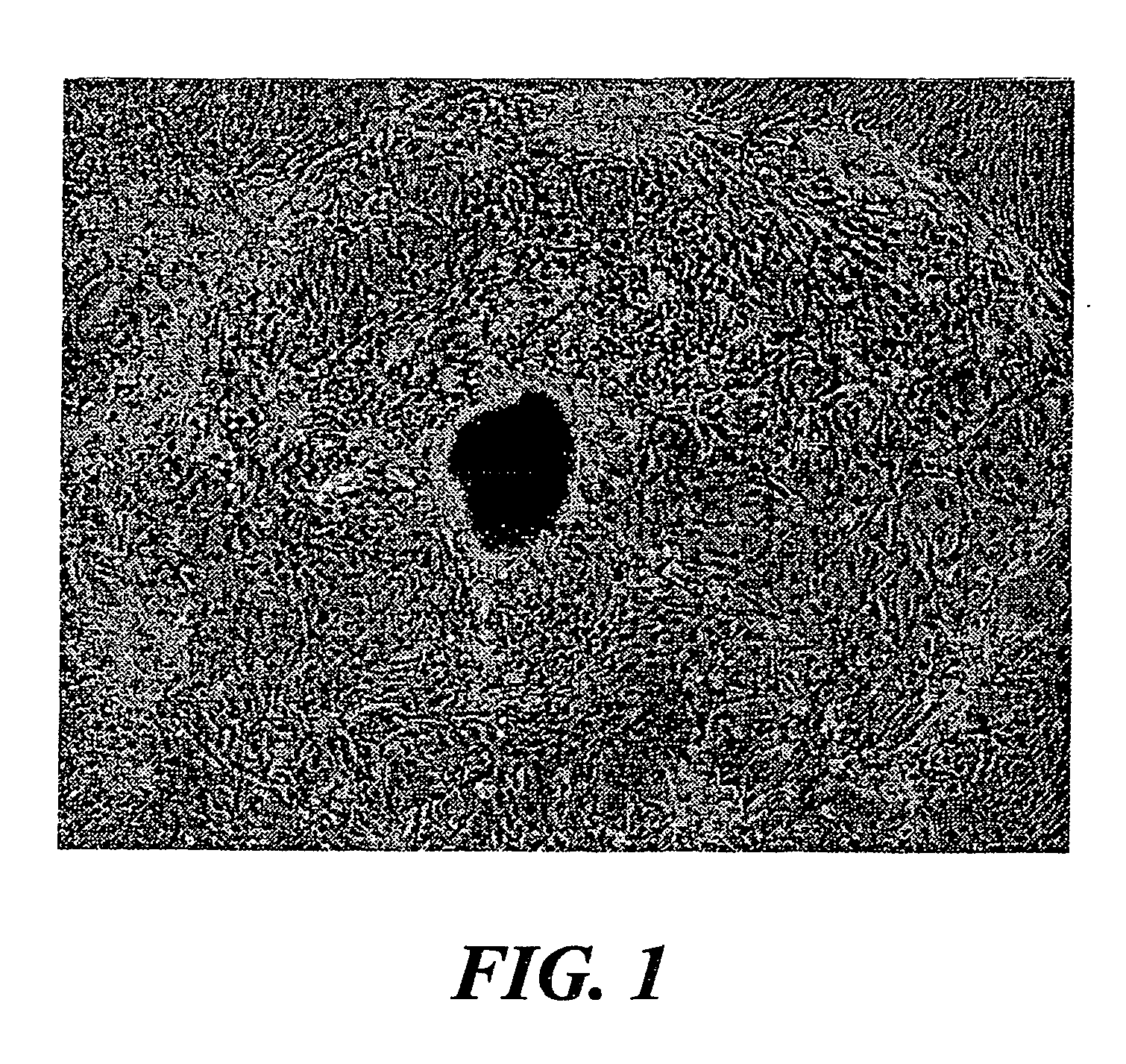
[0056] As early as 24 hours after establishment of the organ culture, adherent cells were visible around most sides of the tissue plug. By 36-48 hours, cells displaying a cobblestone morphology was observed. Within 7-10 days of culture, evenly spaced nodules was observed, which formed a circular pattern around the plug of conjunctival tissue (FIG. 1). Initial attempts at locating goblet cells within this mixed population of cells utilized AB / PAS (a well-documented stain for glycoconjugates in fixed tissue sections). The nodules depicted in FIG. 1 reacted strongly to AB / PAS displaying a dark blue stain indicative of acidic mucin. To further purify potential goblet cells, one nodule was grown in the culture dish. All other cells were scraped from the surface of the vessel using a rubber policeman and the nodule was thoroughly rinsed to rid the culture of floating, non-goblet cells. The nodule was refed with RPMI-1640 medium supplemented...
example ii
Proliferation of Goblet Cells In Vitro
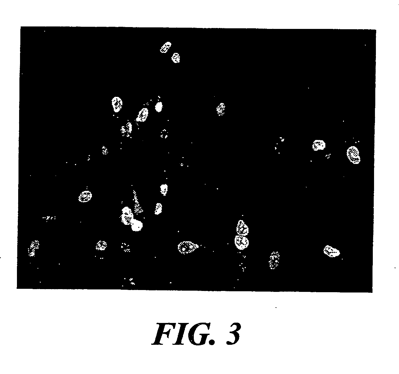
[0058] The proliferation profile of the goblet cell cultures was assessed by staining the cells with an antibody against Ki-67 antigen, a nuclear and nucleolar protein which is exclusively expressed in proliferating cells. (Gerdes et al., 1983; Gerdes et al., 1984) All cultures were routinely evaluated for Ki-67 reactivity. Ki-67 is localized in all primary and passaged cultures of conjunctival goblet cells indicating that our cells are actively proliferating in vitro. As shown in FIG. 3, greater than 30% of the goblet cells in this primary culture were actively proliferating as indicated by their reactivity to Ki-67. During the course of these studies, it was observed that the number of Ki-67 positive cells correlated with the degree of confluence of any given culture. As the cells approached confluence, the number of proliferating cells declined both in primary and passaged cultures.
example iii
Characterization of Cultured Goblet Cells
[0059] Although AB / PAS was used as a screening mechanism to aid in the identification and subsequent purification of the goblet cell cultures, it was important to determine whether or not these purified cells retained positive reactivity to AB / PAS. The present results show that both primary and passaged cultures react histochemically with the stain. Shown in FIGS. 4A (rat) and 13A (human) are cells grown from a representative primary culture in which the cell nuclei are stained blue and displayed a red vesicle-filled cytoplasm. Further staining examined the reactivity of goblet cells, which appeared to contain secretory droplets on their surface. These cells (FIG. 4B) stained a reddish color with no visible demarcation of the nucleus. Droplets located on top of the cells were bright red, indicating that these cells were associated with a basic type of mucin secretion product. Often, cultures appear covered with what appeared to be a fibrous ...
PUM
| Property | Measurement | Unit |
|---|---|---|
| temperature | aaaaa | aaaaa |
| pH | aaaaa | aaaaa |
| time | aaaaa | aaaaa |
Abstract
Description
Claims
Application Information
 - R&D
- Intellectual Property
- Life Sciences
- Materials
- Tech Scout
- Unparalleled Data Quality
- Higher Quality Content
- 60% Fewer Hallucinations
Browse by: Latest US Patents, China's latest patents, Technical Efficacy Thesaurus, Application Domain, Technology Topic, Popular Technical Reports.
© 2025 PatSnap. All rights reserved.Legal|Privacy policy|Modern Slavery Act Transparency Statement|Sitemap|About US| Contact US: help@patsnap.com