
Apparatus, method, and program for producing subtraction images

a subtraction image and apparatus technology, applied in the field of apparatus, method and program for producing subtraction images, can solve the problems of difficult to find a difference (a lesion or the like) between the two images, difficult to superimpose the image on the other, and expensive three-dimensional transmitted images, etc., to achieve accurate detection of abnormal patterns

Inactive Publication Date: 2005-11-24
FUJIFILM HLDG CORP +1
View PDF49 Cites 16 Cited by
  • Summary
  • Abstract
  • Description
  • Claims
  • Application Information

AI Technical Summary

Benefits of technology

[0010] In view of the foregoing circumstances, it is an object of the present invention to provide a subtraction image production apparatus, method and program for accurately detecting abnormal patterns or the like.
[0050] Further, if a low-frequency component is removed from each of the reconstructed cross-sectional images, noise generated during the reconstruction can be eliminated.
[0051] Further, an error due to a change in the posture of the subject may be reduced by superimposing an ordinary radiographic image obtained by radiographing a subject by ordinary radiographic photography on each of cross-sectional images reconstructed from radiographic images obtained by radiographing the subject by tomosynthesis, producing an addition image by adding the cross-sectional images, and further producing a subtraction image between the addition image and the ordinary radiographic image.
[0052] Further, if the past tomosynthesis images are images obtained before the subject was dosed with a contrast medium, and the current tomosynthesis images are images obtained after the subject was dosed with the contrast medium, even if the density of the contrast medium is low, the area in which the contrast medium is photographed is emphasized. Further, since the subtraction image is obtained, surrounding structural elements can be removed. Therefore, the area in which the contrast medium was photographed is emphasized. Alternatively, one of the past images and the current image may be ordinary radiographic images and the other may be tomosynthesis images. In that case, a similar effect can be also achieved.
[0054] Further, a contrast medium photographed area can be automatically detected in each subtraction image. Therefore, an area in which the contrast medium was photographed can be easily recognized.

Problems solved by technology

However, since the three-dimensional transmitted images are expensive, radiologists obtain the three-dimensional transmitted images only at the first examination of a patient, and they obtain two-dimensional transmitted images, which are relatively inexpensive, at later examinations of the patient to monitor a temporal change in the diseased area of the patient.
Therefore, it is difficult to find a difference (a lesion or the like) between the two images in some cases.
Therefore, it is difficult to superimpose the image on the other so that all of the corresponding structural elements of the subject are aligned at the same time.
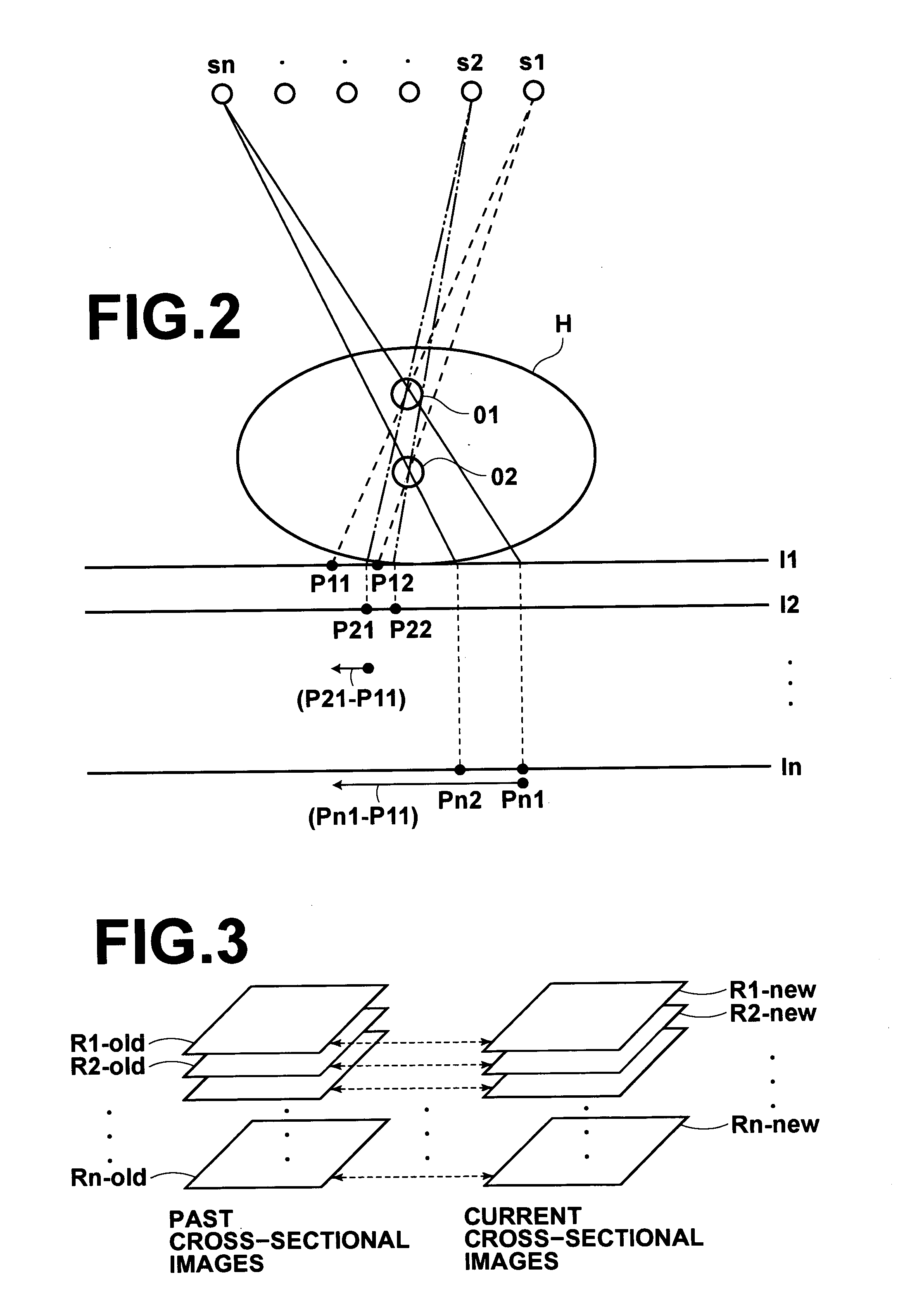
However, when the two-dimensional transmitted images are compared with each other, it is impossible to know at which cross-sectional plane the change is present.

Method used


Image

  • Apparatus, method, and program for producing subtraction images
  • Apparatus, method, and program for producing subtraction images
  • Apparatus, method, and program for producing subtraction images

Examples


Embodiment Construction

[0073] A first embodiment of a subtraction image production apparatus according to the present invention will be described with reference to the attached drawings.

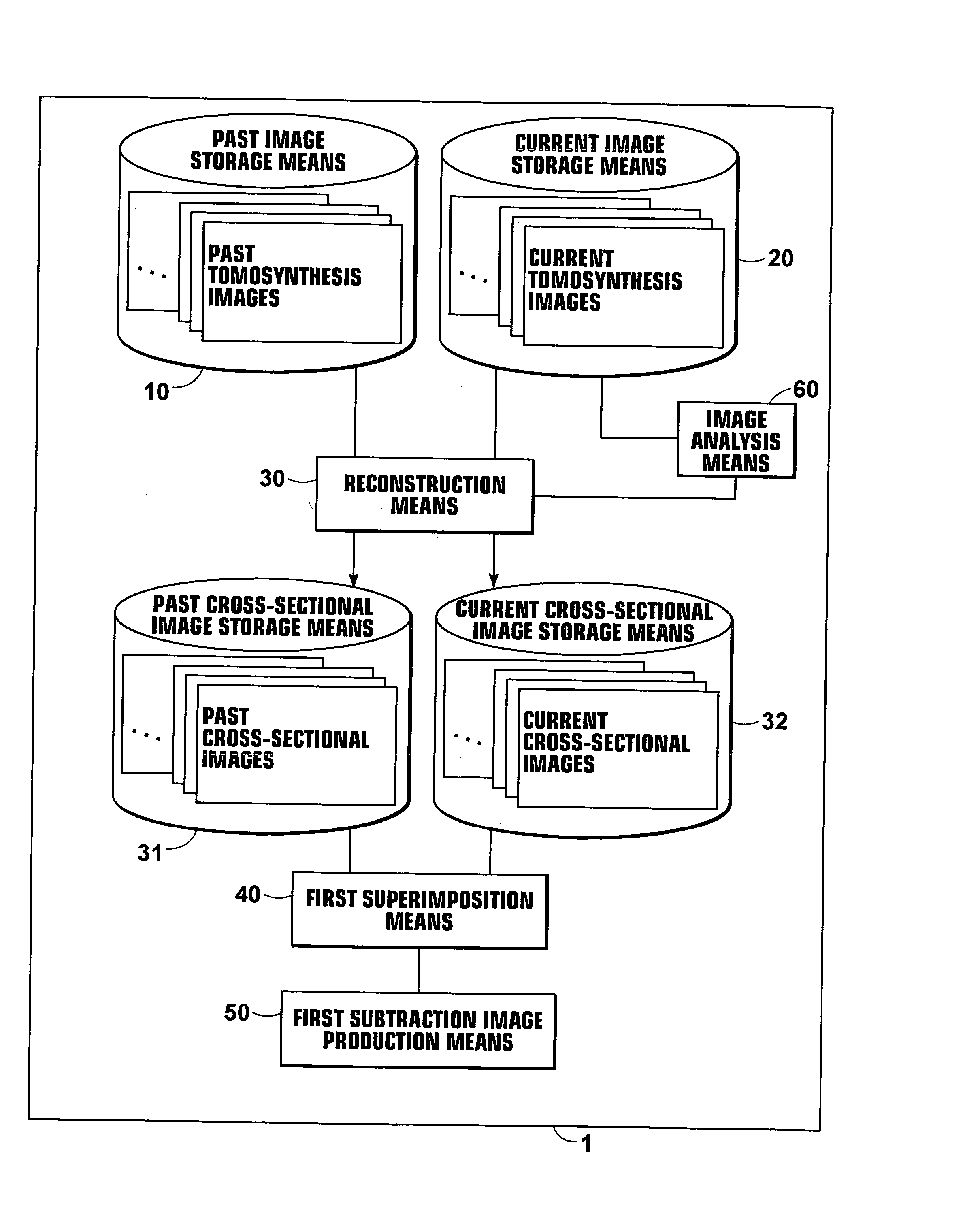
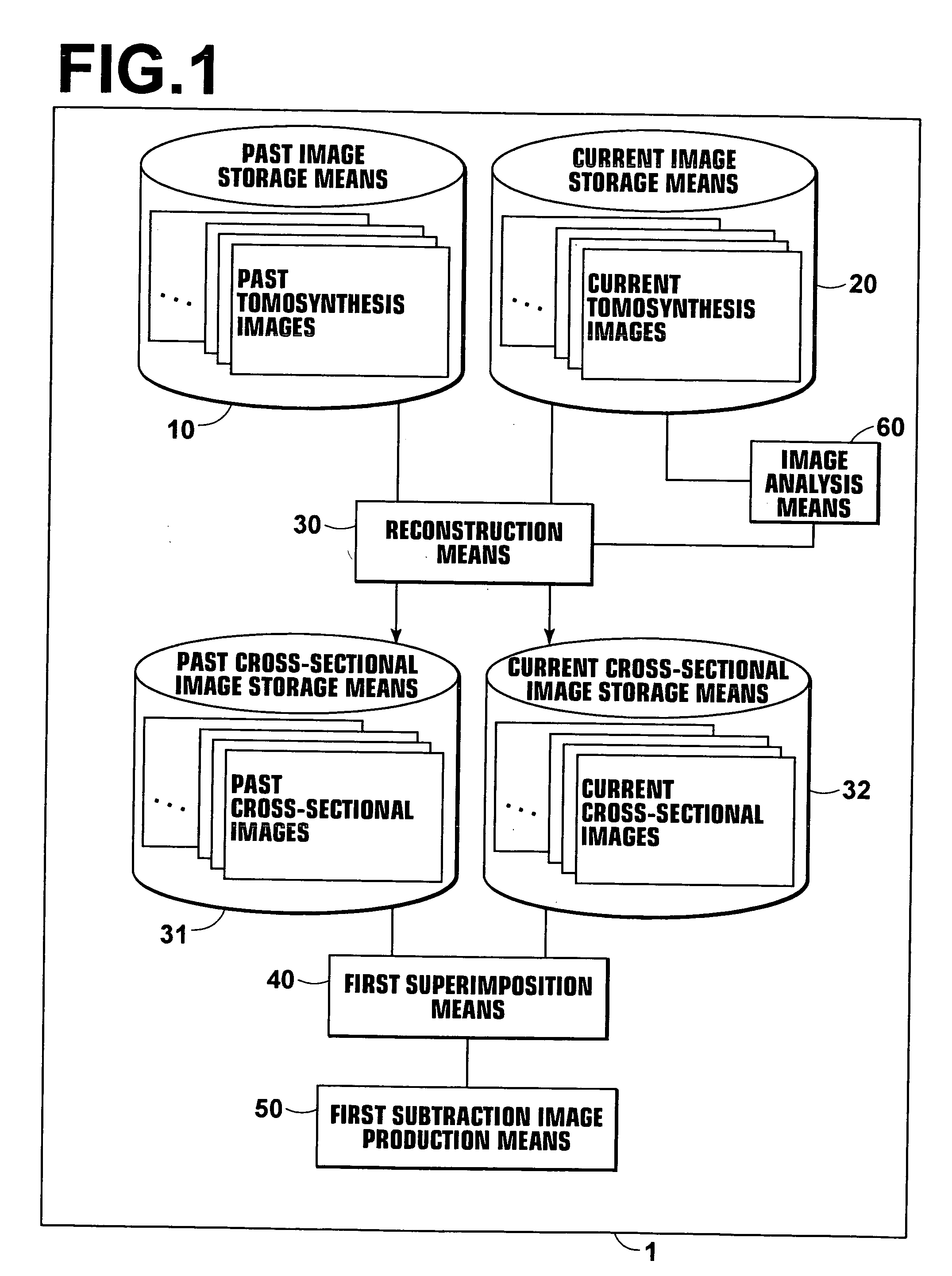
[0074] As illustrated in FIG. 1, a subtraction image production apparatus 1 according to the present invention includes a past image storage means 10 for storing past tomosynthesis images obtained by radiographing a subject by tomosynthesis. The subtraction image production apparatus 1 also includes a current image storage means 20 for storing current tomosynthesis images obtained by radiographing the subject by tomosynthesis. The subtraction image production apparatus 1 also includes a reconstruction means30 for reconstructing cross-sectional images from the tomosynthesis images. The subtraction image production apparatus 1 also includes a first superimposition means 40 for superimposing a past cross-sectional image on a corresponding current cross-sectional image for each cross-sectional plane so as to align structural ...


PUM


Abstract

A plurality of past cross-sectional images are reconstructed from a plurality of past tomosynthesis images obtained by radiographing a subject by tomosynthesis. A plurality of current cross-sectional images are reconstructed from a plurality of current tomosynthesis images obtained by radiographing the subject by tomosynthesis. Each of the plurality of past cross-sectional images is superimposed on a corresponding current cross-sectional image included in the plurality of current cross-sectional images for each cross-sectional plane so as to align structural elements therein. When the structural elements are aligned, a subtraction image between the corresponding past cross-sectional image and current cross-sectional image is produced.

Description

BACKGROUND OF THE INVENTION [0001] 1. Field of the Invention [0002] The present invention relates to an apparatus, method, and program for producing subtraction images from images obtained by X-raying a subject. [0003] 2. Description of the Related Art [0004] Conventionally, a temporal subtraction technique has been proposed to monitor a temporal change in a diseased area of a patient, pictured in a radiographic image. In the temporal subtraction technique, a subtraction image between temporally sequential radiographic images is produced, and a region of the radiographic image, at which the temporal change is present, is emphasized so that radiologists can easily observe the change. It is a diagnosis supporting technique for supporting the radiologists by enabling them to observe the produced subtraction image and the temporally sequential radiographic images at the same time (for example, “Digital Image Subtraction of Temporally Sequential Chest Images for Detection of Interval Cha...

Claims


Application Information

Patent Timeline
IPC IPC(8): A61B6/00A61B6/02G01N23/00G06K9/32G06T5/50G21K1/12H05G1/60
CPCA61B6/025G06T5/50G06T2200/04A61B6/4291G06T2207/20224G06T2207/30061G06T2207/30096G06T2207/10112
Inventor SENDAI, TOMONARI
Owner FUJIFILM HLDG CORP
Learn More