
System and method for analyzing and displaying computed tomography data

a computed tomography and data technology, applied in the field of systems, can solve the problems of affecting colorectal screening and preventive efforts, affecting the detection of colorectal cancer,

Inactive Publication Date: 2005-11-03
NETKISR
View PDF25 Cites 118 Cited by
  • Summary
  • Abstract
  • Description
  • Claims
  • Application Information

AI Technical Summary

Problems solved by technology

Unfortunately, colon cancer also causes approximately 60,000 deaths annually, with only lung cancer causing more deaths among the population of America.
What makes these statistics truly unfortunate is that colon cancers are perhaps one of the most preventable of cancers because they usually begin as benign polyps which grow slowly for five to ten years before becoming cancerous.
Regrettably, widespread colorectal screening and preventive efforts are hampered by several practical impediments, including limited resources, methodologic inadequacies, and poor patient acceptance leading to poor compliance.
In addition, a fecal occult blood test (FOBT) fails to detect the majority of cancers and pre-cancerous polyps.
The accuracy of barium enema varies among centers and is therefore not always reliable.
However, direct interpretation of the cross-sectional images is difficult because a scan of the colon consists of several hundred axial tomograms.
Without advanced image manipulation tools, interpretation of the colon's complex three-dimensional shape from these cross sections alone is very difficult for a medical practitioner.
While these techniques have advanced the art of using CT scanning in assisting the diagnosis of colorectal cancers, among other diseases, the prior techniques typically required massive computing capacity and specialized work stations.
Moreover, the prior techniques have also been limited to providing views of the colorectal pathway that may occlude the presence of polyps, and which do not provide any information regarding the subsurface morphology of the colorectal pathway, or other tubular structure being analyzed.
However, this “unfolding” technique only allows a view of the internal structure of the tubular structure or colon, and does not provide a truly “flattened” view of the internal wall of the colon, wherein the inherent curvature of the colon is “flattened”, thus allowing structures that protrude from the inner wall of the colon, such as Haustral ridges, to hide structures such as small polyps or other anomalies from the practitioners view.
Moreover, none of the systems, methods or techniques currently available are believed to be able to provide an assessment of the wall of the tubular structure.
Such cancers cannot be observed using the means described above, since none of those methods allow for an analysis of the wall thickness or density of the tubular structure.

Method used

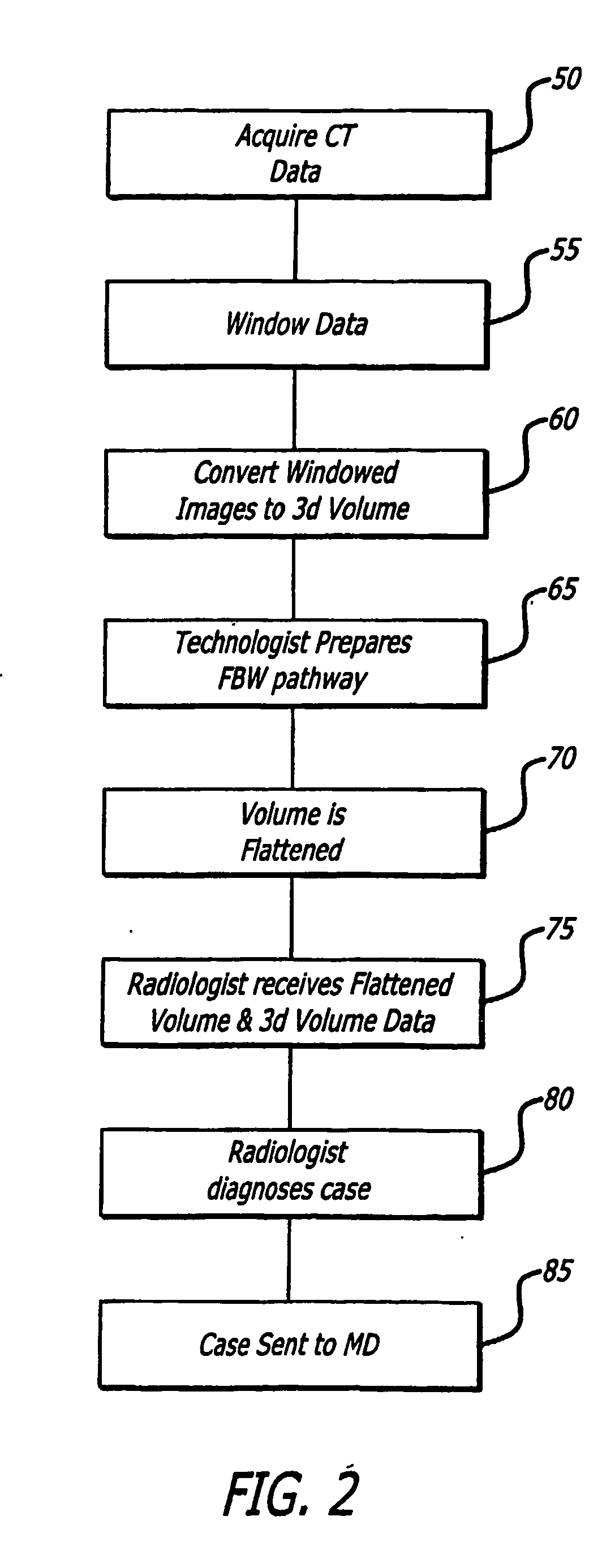

Image

  • System and method for analyzing and displaying computed tomography data
  • System and method for analyzing and displaying computed tomography data
  • System and method for analyzing and displaying computed tomography data

Examples


Embodiment Construction

[0054] The present invention generally relates to a method and system for generating and displaying interactive two- and three-dimensional figures representative of structures of the human body. The three-dimensional structures are in the general form of selected regions of the body and, in particular, body organs with hollow lumens such as colons, tracheobronchial airways, blood vessels, and the like. In accordance with the method and system of the present invention, interactive, three-dimensional renderings of a selected body organ are generated from a series of acquired two-dimensional images generated from data acquired by a computerized tomographic scanner, or CAT scan. Recent advances in CAT scanning technology, notably the advent of use of helical or spiral CAT scanners, have revolutionized scanning of human body structures because they allow the collection of a vast amount of data in a relatively short period of time. For example, an entire colorectal scan may be accomplishe...


PUM


Abstract

A system (10) and method are provided for analyzing and displaying images rendered from data sets resulting from a scan of a patient. The images displayed include both two-dimensional and three-dimensional views of selected portions of a patient's anatomy, for example, a tubular structure such as a colon.

Description

CROSS-REFERENCE TO RELATED APPLICATION [0001] This application claims the benefit of priority to U.S. Provisional Applications No. 60 / 364,298, filed Mar. 14, 2002 and U.S. Provisional Applications No. 60 / 429,827, filed Nov. 27, 2002, the subject matter of each being incorporated herein in its entirety.BACKGROUND OF THE INVENTION [0002] 1. Field of the Invention [0003] The present invention generally relates to analysis and display of data acquired using computed tomography representing various organs of the human body. More specifically, the present invention relates to a computerized system for analyzing data acquired from tubular structures of the body, for example, the colon or gastro-intestinal tract, and displaying that data in a manner providing for rapid evaluation of the tissue structures of tubular structure and the accurate determination of any abnormalities present. [0004] 2. Description of Related Art [0005] In industrialized nations, colorectal cancer is the second lead...

Claims


Application Information

Patent Timeline
IPC IPC(8): A61B6/03A61B5/05A61B5/055A61B6/00G06F19/00G06T15/08
CPCA61B5/055A61B5/4255A61B6/03A61B6/032A61B6/481G06T2219/028G06F19/3437G06T15/08G06T19/00G06T2210/41G06T2219/021G06F19/321G16H50/50G16H30/40G16H30/20Y02A90/10
Inventor GLENN JR., WILLIAM V.KEMPER, ROGER
Owner NETKISR
Learn More