
Device and method of detecting mutations and polymorphisms in DNA

Inactive Publication Date: 2005-07-28
AGENCY FOR SCI TECH & RES +1
View PDF13 Cites 60 Cited by
  • Summary
  • Abstract
  • Description
  • Claims
  • Application Information

AI Technical Summary

Benefits of technology

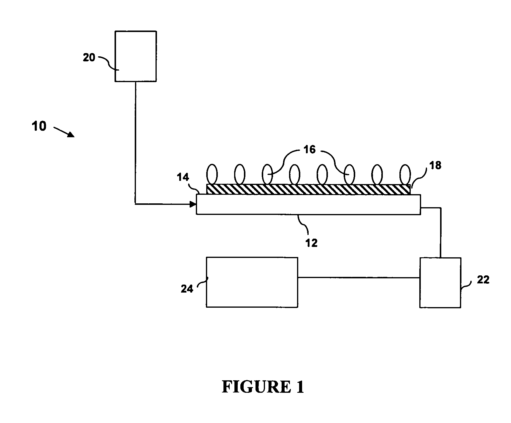
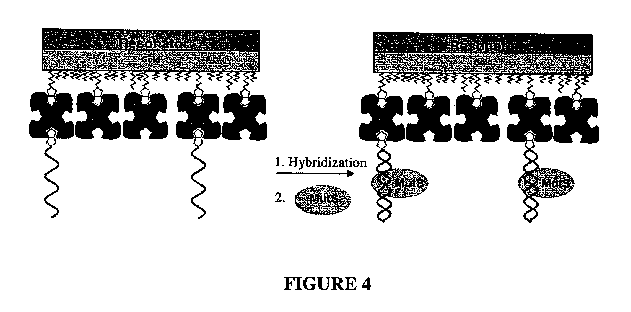
[0008] The present invention provides a resonator sensor having an immobilized capture molecule, either probe DNA containing non-disease DNA sequence, or a mismatch binding molecule. The resonator sensors described herein are useful for detection of a mutation or a polymorphism in target DNA. The present methods are advantageous in that they are simple, requiring relatively simple instrumentation, and do not involve gel electrophoresis or staining techniques.
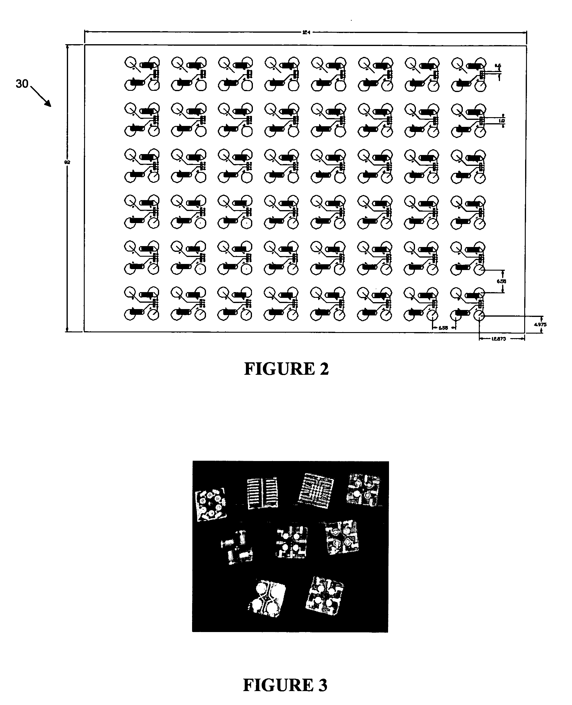
[0010] Furthermore, as resonator sensors are easy to miniaturize and automate, the present methods may be adapted for resonator array techniques, for high throughput and rapid screening of multiple DNA samples or multiple mutations at one time.

Problems solved by technology

These methods typically involve time-consuming or technically complicated methods that are not suitable for handling a large number of samples at one time, such as gel-electrophoresis and / or staining procedure.
Some of the methods require that the exact location of the mutation be known, and the results can be difficult to interpret when the target DNA is heterozygous for the mutation in question.
Thus, such techniques are not practical for use in screening of large numbers of samples for polymorphisms.
These methods, however, are unsuitable for scanning wide regions of DNA (a capability essential for the detection of genomic polymorphisms) as single base substitutions or deletions result in very small differences in the hybridization affinity.
Since these methods use chromatography methods, they are not well adapted to parallel analysis of multiple samples.

Method used


Image

  • Device and method of detecting mutations and polymorphisms in DNA
  • Device and method of detecting mutations and polymorphisms in DNA
  • Device and method of detecting mutations and polymorphisms in DNA

Examples


example 1

[0111] Detection of G:T Mismatch using Method I: FIG. 6 shows the frequency response of a QCM to a whole reaction procedure using the method depicted in FIG. 4, starting from a biotin-thiol treated surface.

[0112] Briefly, streptavidin in PBS buffer (0.1 mg / ml) was immobilized on a surface which was first reacted with a biotin-thiol molecule based on biotin-streptavidin interaction. BSA was then applied (5 mg / ml in PBS) to block any possible free gold surface that might be present due to the low biotin self-assembled monolayer coverage. The baseline was determined using circulated Tris-HCl buffer (20 mM Tris-HCl, pH 7.5, 200 mM NaOH, 1 mM DTT, 0.1 mM EDTA, and 5 mM MgCl2), followed by MutS buffer blank alone, which contains the same amount of glycerol and Triton™ X-100 as in the MutS protein sample, which further contains MutS at a final concentration of 100 nM (diluted 1:200 from the stock buffer, using Tris-HCl buffer). The subsequent application of the MutS protein produced a bar...

example 2

[0118] Detection of 1 to 4 Deletions: In this example, probe 4 was immobilized and target DNAs (SEQ ID NOS.:5 to 8) (1 uM) were hybridized to form heteroduplexes containing 1 to 4 unpaired bases. Again, the hybridization profile between the MM0 and unpaired DNA showed no significant difference, while the subsequent MutS binding shows obvious difference in both the binding amount and binding kinetics. The consequent response frequency upon MutS binding these DNA was 92 Hz (unpaired CA), 90 Hz (unpaired C), 76 Hz (unpaired CAG), and 70 Hz (unpaired CAGG), which indicates that heteroduplexes containing 1 to 4 unpaired bases were detectable, but not equally well. This is likely due to differing binding affinities of MutS, resulting in different saturation amounts of MutS on each heteroduplex. The order of the detection sensitivity appeared to be 2 unpaired bases≧1 unpaired base>3 unpaired bases>4 unpaired bases. For the heteroduplex with 3 or 4 unpaired bases, the MutS binding was less ...

example 3

[0119] Motional Resistance Measurement of MutS Binding: In this experiment, the QCM sensor carrying a DNA heteroduplex containing two unpaired bases, CA, was connected to an S&A 250B Network Analyzer (Saunders & Associates, Inc., USA). The oscillation spectrum of the QCM before and after MutS binding was recorded (FIG. 10). FIG. 11 shows the kinetic curves of the frequency (A) and motional resistance (B) changes upon MutS binding on the unpaired CA DNA and a homoduplex DNA. The ratio between ΔR and ΔF for the MM0 (0.033) was greater than that for the unpaired CA (0.025). This difference is believed to relate to the different nature of the MutS binding to homoduplex DNA as compared to heteroduplex DNA. The adsorbed protein based on non-specific contact with the MM0 DNA backbone tends to be more flexible when compare to that based on unpaired bases recognition.


PUM

PropertyMeasurementUnit
Frequencyaaaaaaaaaa
Energyaaaaaaaaaa
Affinityaaaaaaaaaa

Abstract

There is provided a resonator sensor useful for detecting polymorphisms and mutations in DNA. The resonator sensor has a capture molecule immobilised on its surface, the capture molecule being either a probe DNA containing a reference sequence, or a mismatch binding molecule, and being capable of forming a probe DNA / target DNA / mismatch binding molecule complex on the surface of the resonator. A method for detecting mutations in a target DNA, including single nucleotide polymorphisms, is also provided.

Description

CROSS-REFERENCE TO RELATED APPLICATIONS [0001] The present application claims priority from U.S. provisional patent application No. 60 / 504,434, filed on Sep. 22, 2003 and U.S. provisional patent application No. 60 / 534,366, filed on Jan. 6, 2004, the contents of which are hereby incorporated herein by reference.FIELD OF THE INVENTION [0002] The present invention relates generally to devices and methods for detection of mutations and polymorphisms in DNA, and particularly to use of mismatch binding proteins and ligands for same. BACKGROUND OF THE INVENTION [0003] A polymorphism is a variation in genetic sequence in which an individual's genetic sequence at a given location in the genome varies from the sequence commonly found in a population. Polymorphisms can be induced by mutagens, or they may be naturally occurring in a population. Presence of a polymorphism in an individual's DNA can often be used as a disease marker, since particular polymorphisms tend to be linked to certain dis...

Claims


Application Information

Patent Timeline
IPC IPC(8): C12Q1/68G01N33/48G01N33/50G01N33/543G06F19/00
CPCC12Q1/6825C12Q2600/156G01N33/54373C12Q2565/607
Inventor SU, XIAODIROBELEK, RUDOLFKNOLL, WOLFGANGO'SHEA, SEAN
Owner AGENCY FOR SCI TECH & RES
Learn More