
Tibia condyle resurfacing prosthesis

a technology for resurfacing prostheses and tibias, which is applied in the field of tibia condyle resurfacing prosthesis, can solve the problems of low bone wear when compared to metals or ceramics, and achieve the effect of low friction and low abrasion

Pending Publication Date: 2022-06-09
KYON
View PDF0 Cites 0 Cited by
  • Summary
  • Abstract
  • Description
  • Claims
  • Application Information

AI Technical Summary

Benefits of technology

The present invention solves issues in veterinary medicine with a coated implant that can be used to resurface only one side of the knee joint. The implant is a dish-shaped dagger that mimics the natural shape of the knee joint. It is made with a low friction coating that reduces wear and tear on the bone. The femoral condyle slides into the implant's concave recess, providing stability to the joint. The bone-facing surface of the implant is coated for bony integration. The technique allows for easy access for fixation of the implant while preserving the insertion of the medial collateral ligament.

Problems solved by technology

They also result in very low wear of bone when compared to metals or ceramics.

Method used


Image

  • Tibia condyle resurfacing prosthesis
  • Tibia condyle resurfacing prosthesis
  • Tibia condyle resurfacing prosthesis

Examples


Embodiment Construction

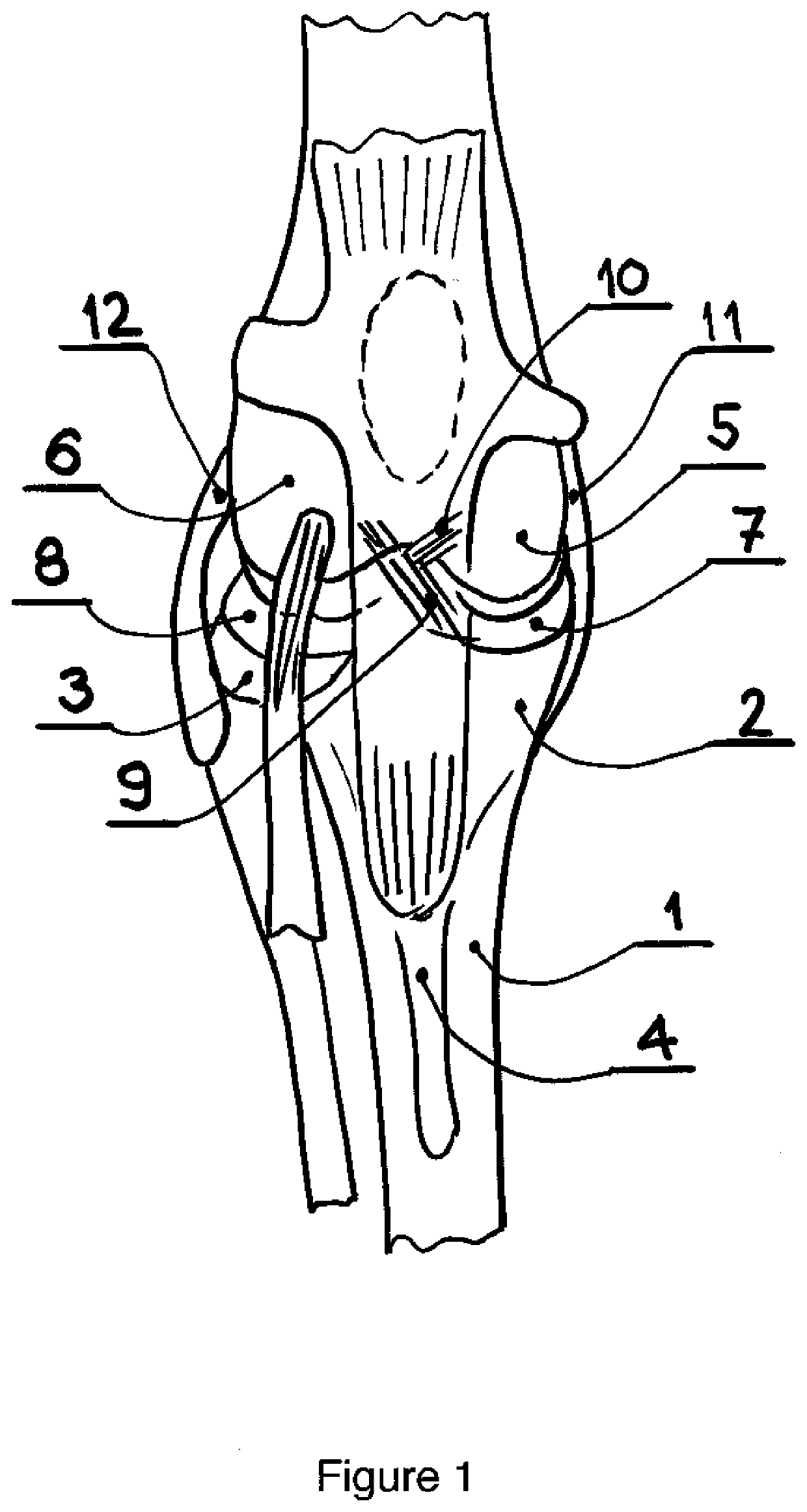
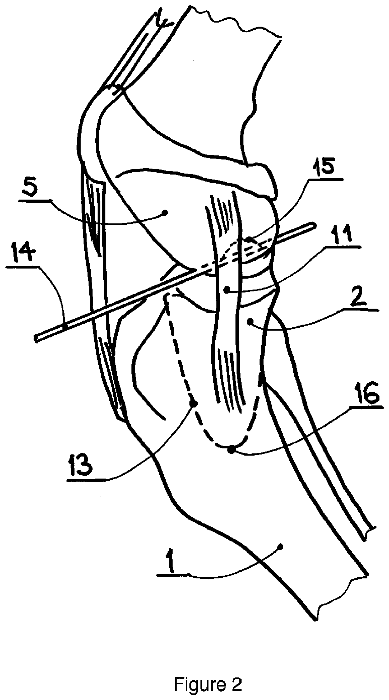
[0024]A frontal perspective view of the dog stifle (knee), FIG. 1, illustrates basic anatomical features relevant to this invention. The proximal aspect of the tibia 1 flares out laterally and medially, creating a broad plateau, with the medial condyle 2 and the lateral condyle 3. Cranially to the plateau, the bone is narrowed to form the tibial tuberosity 4. Stifle joint comprises medial 5 and lateral condyles 6 of the femur, which articulate with respective condyles of the tibia. Interposed between the condyles of the femur and the tibia are menisci 7 and 8. With two bones of convex shapes, the joint is inherently unstable. Ligaments that span the joint prevent luxation. There are two main pairs: (i) cruciate ligaments 9 and 10 within the joint prevent dislocation by translation in the sagittal plane and limit the internal rotation of the tibia; (ii) collateral ligaments 11 and 12 prevent varus-valgus angulation in the frontal plane and also limit the external rotation of the tibi...


PUM

PropertyMeasurementUnit
shapeaaaaaaaaaa
stabilityaaaaaaaaaa
frictionaaaaaaaaaa

Abstract

The present invention resolves the main problems of bone-sparing resurfacing surgery of the medial compartment of the knee in dogs and in people. With a hard, low abrasion, low friction coating of the implant (100), such as ADLC or pyrolytic carbon, it is possible to resurface only one side of the joint. In this case, the tibia plateau resurfacing with a concave recess on its femur-facing surface provides not only pain-free mobility but also the necessary stability of the joint with a deficient cruciate ligament. The planar osteotomy of the medial condyle, with or without resurfacing, can also be used to emulate TPLO procedure limited to the medial compartment of the stifle.

Description

[0001]The present invention relates to a medial tibial condyle resurfacing implant for hemiarthroplasty of the knee joint, for use in human and veterinary medicine. Further, the present invention relates to a medial tibial condyle osteotomy emulating procedure for stabilization of the cranial cruciate deficient stifle in human and animal knees.BACKGROUND[0002]In people but also in dogs, the medial condyles of the tibia and the femur are at high risk of degenerative arthritis as among other possible causes a consequence of cruciate ligament rupture or of skeletal deformities resulting in increased loading of the medial knee compartment.[0003]In human orthopedics, the usual approach is to perform either a total knee replacement or, increasingly, to replace only smaller areas of the affected joint.[0004]In human orthopedics, the medial compartment of the knee is also commonly affected by overloading due to various deformity of the leg. If the cartilage is still able to support the load...

Claims


Application Information

Patent Timeline
Patent Type & Authority Applications(United States)
IPC IPC(8): A61F2/38
CPCA61F2/389A61F2002/307A61F2002/3895A61F2002/30769A61F2310/00574A61F2310/0058
Inventor TEPIC, SLOBODANBRESINA, STEPHEN
Owner KYON
Learn More