Purification of the leading front of migratory cells
a technology of migratory cells and purification methods, applied in the field of purification of the leading front of migratory cells, can solve the problems of poor chemoattractant for both cell types, inability to specifically isolate the pseudopodium and the cell body for biochemical comparison using previous techniques, and lack of cellular material available for analysis
- Summary
- Abstract
- Description
- Claims
- Application Information
AI Technical Summary
Problems solved by technology
Method used
Image
Examples
example 1
[0073] Quantitative Pseudopodia Assay
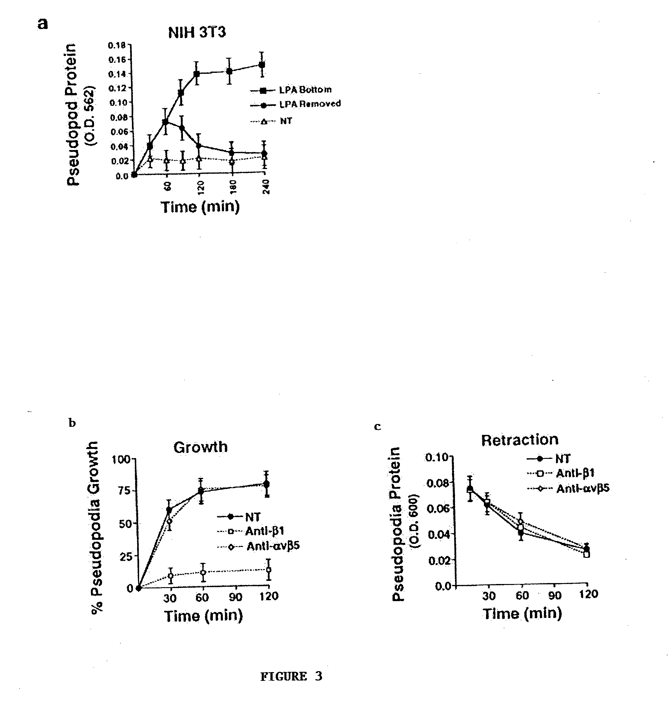
[0074] Pseudopodia extension was monitored using a pseudopodia assay kit (ECM 650; Chemicon International) or Costar chambers. Serum-starved cells (75,000) were placed into the upper compartment of a chamber (6.5 .mu.m) equipped with a 3.0-.mu.m porous polycarbonate membrane coated on both sides with an optimal amount of ECM protein (5 .mu.g / ml fibronectin or collagen type I). Cells were allowed to attach and spread on the upper surface of the membrane for 2 h, and then stimulated with LPA (Sigma-Aldrich), insulin, or buffer only, which was placed in the lower chamber to establish a gradient or placed in the upper and lower chamber to form a uniform concentration. Cells were allowed to extend pseudopodia through the pores toward the direction of the gradient for various times. To initiate pseudopodia retraction, the chemoattractant was removed or an equivalent amount of chemoattractant placed in the upper chamber to create a uniform concentration...
example 2
[0075] LPA and Insulin as Chemoattractants
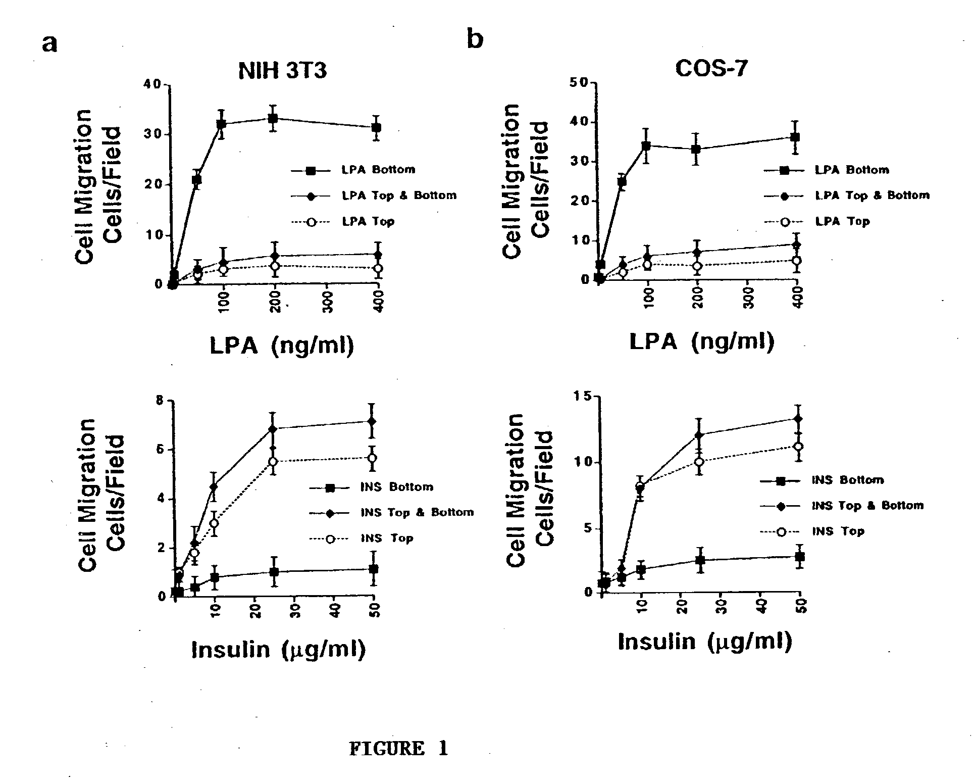
[0076] NIH 3T3 cells were examined for cell migration for 3 h in 8.0-.mu.m porous Boyden chambers containing different concentrations of LPA or insulin placed in 1) the bottom, 2) top, or 3) top and bottom compartments. The number of migratory cells per microscopic field (200.times.) on the underside of the membrane was counted as described in Example 1 above. Additionally, COS-7 cells were examined for cell migration as described above with respect to NIH 3T3 cells. The results are set forth in FIG. 1, where it is seen that LSA is a potent chemoattractant for both cell types. In FIG. 1, each point on the graphs represents the mean.+-.SEM of three triplicate migration chambers of three independent experiments.
example 3
[0077] Pseudopodia Extension
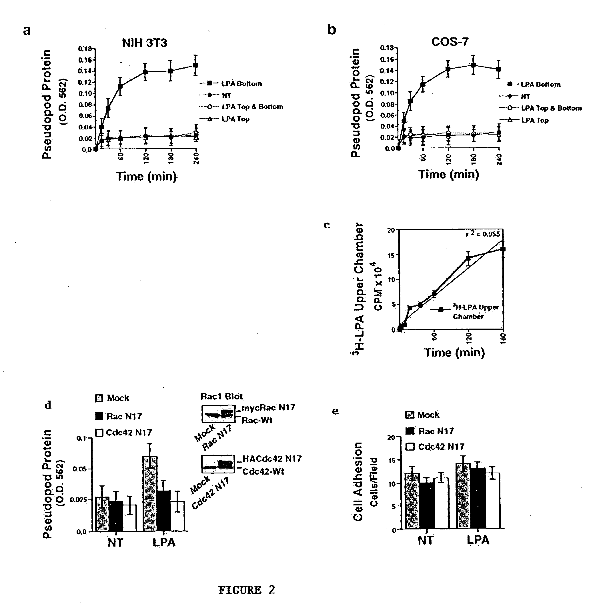
[0078] NIH 3T3 cells were allowed to attach to fibronectin coated 3.0-.mu.m porous membranes for 2 h. Pseudopodia extension was then examined for various times in the absence (NT) or presence of LPA (100 ng / ml) in the bottom, top, or top and bottom compartments. Pseudopodia protein on the underside of the membrane was determined as described in Example 1. Results are set forth in FIG. 2, where each point in the graphs represents the mean.+-.SEM of three triplicate membranes of three independent experiments. COS-7 cells were examined for pseudopodia protrusion in the same manner as for NIH 3T3 cells as described above.
[0079] Confocal images of NIH 3T3 pseudopodia protrusion on the undersurface of a 3.0-.mu.m porous membrane in response to LPA (100 ng / ml) as a gradient (LPA bottom) or uniform concentration (LPA top and bottom) were generated. Cells were labeled with cell tracker green and then fixed at the indicated times to visualize protruding pseudopodia...
PUM
| Property | Measurement | Unit |
|---|---|---|
| Fraction | aaaaa | aaaaa |
| Adhesivity | aaaaa | aaaaa |
| Pore | aaaaa | aaaaa |
Abstract
Description
Claims
Application Information
 - Generate Ideas
- Intellectual Property
- Life Sciences
- Materials
- Tech Scout
- Unparalleled Data Quality
- Higher Quality Content
- 60% Fewer Hallucinations
Browse by: Latest US Patents, China's latest patents, Technical Efficacy Thesaurus, Application Domain, Technology Topic, Popular Technical Reports.
© 2025 PatSnap. All rights reserved.Legal|Privacy policy|Modern Slavery Act Transparency Statement|Sitemap|About US| Contact US: help@patsnap.com


