
Apparatus and method for treating tumors near the surface of an organ

a radiofrequency electrosurgical and tumor technology, applied in the field of radiofrequency electrosurgical apparatus structure and use, can solve the problems of difficult prediction of the total amount of heat which must be delivered in order to effectively necrose the tissue, large volume ablation and necrosis of highly vascularized tissue, such as liver tissue,

Inactive Publication Date: 2001-08-09
BOSTON SCI SCIMED INC
View PDF2 Cites 67 Cited by
  • Summary
  • Abstract
  • Description
  • Claims
  • Application Information

AI Technical Summary

Benefits of technology

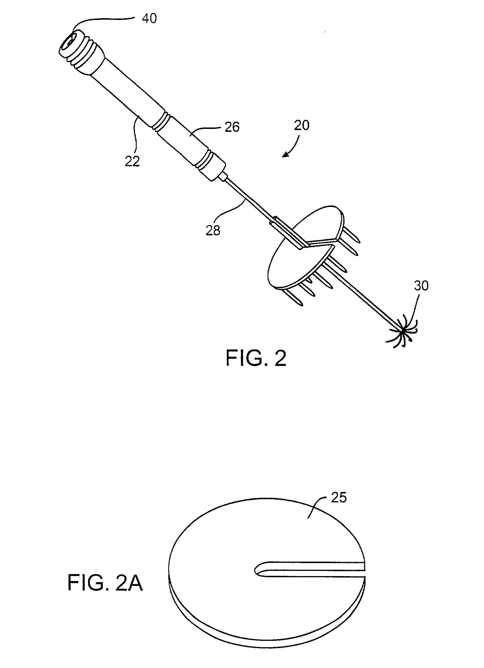
[0012] In a second aspect, the present invention provides for applying electrical energy, such as radiofrequency or other high frequency energy, to an internal tissue site while a cover is deployed over the tissue surface adjacent to the target region within the tissue which is being treated. The cover may comprise electrode(s) as described above. Alternatively, the cover may be electrically neutral (unpowered) and / or electrically insulated to protect surrounding tissue and treating personnel, as described above. Such covers should be capable of compressing the tissue in order to inhibit blood flow to and from the target region being treated. By inhibiting blood flow, energy losses can be minimized and temperature uniformity enhanced. The covers may be rigid plates, conformable surfaces, or the like, and will typically be clipped or otherwise removably or positionably attached to a primary treatment probe. In addition to being electrically insulated, the cover will preferably also be thermally insulating to protect adjacent tissue structures from thermal damage and further to inhibit heat losses from the tissue which is being treated. While in some instances the cover may be discontinuous, i.e., perforated or having other openings or apertures, it will usually be desirable to utilize a cover having a continuous surface which can inhibit the loss of steam from the tissue which is being treated. Containment of steam within the region being treated further enhances tissue heating. Preferably, the compressive force between a deployed electrode array on the primary treatment probe and the cover on the tissue surface will be in the range from 0.5 psi to 1.5 psi, preferably 0.8 psi to 1.2 psi.
[0018] In a still separate aspect of the methods of the present invention, deployment of the first electrode array and second electrode or other cover in a manner such that tissue is compressed therebetween will (after deployment) also inhibit blood flow into and from the target region between the electrodes. Thus, the step of inhibiting blood flow may be achieved as simply as compressing the tissue in order to reduce blood flow through the target region between the electrodes. The compressive forces may be applied by any structure deployed over or otherwise adjacent to the tissue region being treated, usually being positioned directly over the target region in the organ being treated. Typically, the primary electrode which is deployed within the tissue will act as an anchor and the cover or other structure will be secured to a probe or shaft which is part of the electrode. The cover and electrode can then be drawn together and secured in place to compress all or a portion of the tissue volume being treated. In this way, blood flow into and out of the region may be significantly decreased. Optionally, such compression is achieved using treatment electrodes which are also used for introducing a frequency or other electrical current into the treatment region to effect the heating.
[0022] The surface electrode may optionally be connected to the probe using a connector. Usually, the connector will attach the surface electrode in a generally transverse orientation relative to the axis of the probe. Optionally, the connector can be flexible or in the form of a swivel or "universal joint" which permits the surface electrode to align itself with the tissue surface even when the probe is entering at an angle relative to the tissue surface which is not perpendicular. Other suitable connectors include clips, pinch clamps, threaded connectors, hook and loop fasteners (such as VELCRO.TM. brand fasteners), and the like. The connectors should allow the surface electrode or other cover to be selectively attached at various points along the length of the probe. Optionally, the probe can be marked with indicia indicating the depth of penetration, i.e., length between the distal tip of the probe and the particular point on the probe where the surface electrode or other cover may be attached. The ability to connect the surface electrode or other cover to the probe prior to deployment of the first electrode array is useful in helping to position the probe at a desired treatment depth, as described elsewhere herein.

Problems solved by technology

Monopolar treatment is advantageous in permitting formation of two fully formed lesions (necrosed regions) that can be overlapped to treat a desired region, but is disadvantageous since it requires use of a dispersive electrode.
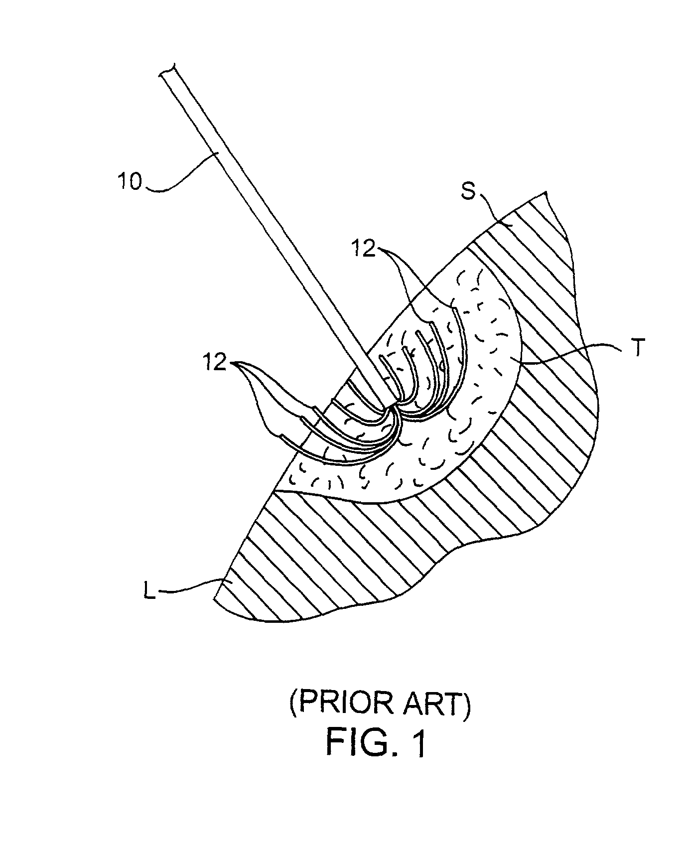
Large volume ablation and necrosis of highly vascularized tissue, such as liver tissue, can be difficult because of thermal transport from the region due to local blood flow.
Moreover, because the degree of vascularization in any particular region is unpredictable, the total amount of heat which must be delivered in order to effectively necrose the tissue is difficult to predict.

Method used


Image

  • Apparatus and method for treating tumors near the surface of an organ
  • Apparatus and method for treating tumors near the surface of an organ
  • Apparatus and method for treating tumors near the surface of an organ

Examples


Embodiment Construction

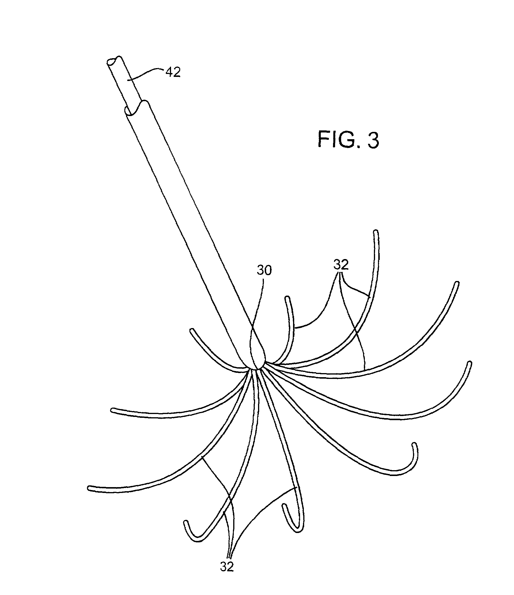
[0035] Systems according to the present invention are designed to position electrode elements and assemblies within and over a treatment region within solid tissue of a patient. The treatment region may be located anywhere in the body where hyperthermic exposure may be beneficial. Most commonly, the treatment region will comprise a solid tumor within an organ of the body, such as the liver, kidney, pancreas, breast, prostate (not accessed via the urethra), and the like. The volume to be treated will depend on the size of the tumor or other lesion, typically having a total volume from 1 cm.sup.3 to 150 cm.sup.3, usually from 1 cm.sup.3 to 50 cm.sup.3, and often from 2 cm.sup.3 to 35 cm.sup.3. The peripheral dimensions of the treatment region may be regular, e.g. spherical or ellipsoidal, but will more usually be irregular. The treatment region may be identified using conventional imaging techniques capable of elucidating a target tissue, e.g. tumor tissue, such as ultrasonic scanning...


PUM


Abstract

A system for treating a target region in tissue beneath a tissue surface comprises a probe for deploying an electrode array within the tissue and a surface electrode for engaging the tissue surface above the treatment site. Preferably, surface electrode includes a plurality of tissue-penetrating elements which advance into the tissue, and the surface electrode is removably attachable to the probe. The tissue may be treated in a monopolar fashion where the electrode array and surface electrode are attached to a common pole on an electrode surgical power supply and powered simultaneously or successively, or in a bipolar fashion where the electrode array and surface electrode are attached to opposite poles of the power supply. The systems are particularly useful for treating tumors and other tissue treatment regions which lie near the surface.

Description

[0001] This application is a continuation-in-part of application Ser. No. 09 / 124,152, filed on Jul. 28, 1998, the full disclosure of which is incorporated herein by reference.[0002] 1. Field of the Invention[0003] The present invention relates generally to the structure and use of radiofrequency electrosurgical apparatus for the treatment of solid tissue. More particularly, the present invention relates to an electrosurgical system having pairs of electrodes and electrode arrays which are deployed to treat large volumes of tissue, particularly for the treatment of tumors which lie close to the surface of an organ.[0004] The delivery of radiofrequency energy to target regions within solid tissue is known for a variety of purposes. Of particular interest to the present invention, radiofrequency energy may be delivered to diseased regions in target tissue for the purpose of causing tissue necrosis. For example, the liver is a common depository for metastases of many primary cancers, su...

Claims


Application Information

Patent Timeline
IPC IPC(8): A61B18/14
CPCA61B18/1477A61B2018/00577A61B2018/143A61B2018/1432
Inventor BEHL, ROBERT S.KERMIT, EBEN L.
Owner BOSTON SCI SCIMED INC
Learn More