Method for processing blood sample
a blood sample and processing method technology, applied in the field of blood sample processing, can solve the problems of difficult detection or obtaining cells found in blood at much lower levels, limited ability of cellsearch system to capture cancer cells, and inability to capture cells by cellsearch system, so as to reduce the filtration area and improve the capture rate of rare cells
- Summary
- Abstract
- Description
- Claims
- Application Information
AI Technical Summary
Benefits of technology
Problems solved by technology
Method used
Image
Examples
example 1
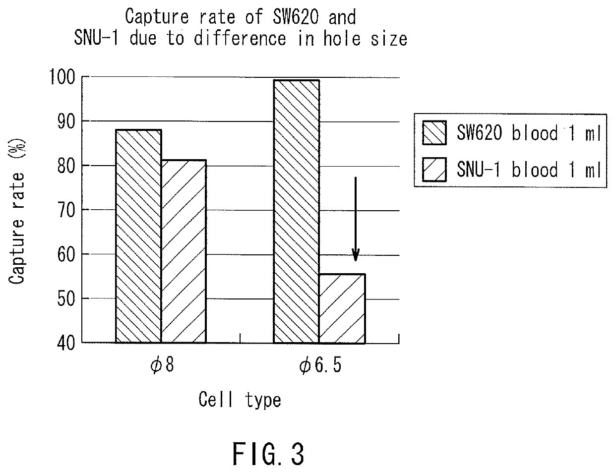
[0146]In the following examples, using filters having the same number of holes and the same filtration area, blood samples were processed in a volume of 14 nl per hole (the total volume of the blood sample to be processed: 1 ml). Consequently, the recovery rate of small cells that would pass through 10 μm holes was generally high as long as the holes were smaller than the cells regardless of the hole shape. On the other hand, the recovery rate of deformable cancer cells was low when the holes were small. Table 3 and FIG. 3 show data comparing the capture capabilities of various holes among the filters. In this case, the total volume of the blood sample to be processed was 1 ml. As shown in Table 3, whether the hole shape was a true circle or an ellipse, the capture rate of deformable cancer cells was significantly reduced as the hole area became smaller. Moreover, clogging (blockage) occurred in ϕ 4 μm holes before the blood sample in a volume of 14 nl / hole had reached the total vol...
example 2
[0148]The amount of a blood sample to be processed through each of the filters was considered. If a large amount of blood can be processed with the same filtration area, it is possible to reduce the filtration area of the filter to be used, to suppress the bulk of the filtration portion in the device, and to ensure cost effectiveness, as compared to the conventional examples. Tables 4 and 5 show average data of the capture rates of SW620 cells and SNU-1 cells, respectively. The average data are also plotted in FIG. 4. In this case, the ϕ 8 filter, the ϕ 9.8×6.5 filter, and the ϕ 12.7×5 filter were used, each of which achieved a better capture rate for 1 ml of blood, and the amount of the blood sample to be processed was increased up to 25 ml. Surprisingly, when the filtration area was 20 mm2, these filters continued to process blood without clogging (blockage) until 25 ml (filtration time: 50 min to 70 min). Thus, a high capture rate of small cancer cells was maintained (see Table 4...
example 3
[0153]Next, data of the capture rates of cells when the filtration pressure was varied during filtration will be shown in the following. In general, high pressure would make it easy for cells to pass through the holes of a filter, and thus degrade the capture capabilities, and low pressure would make it difficult for cells to pass through the holes of a filter, and thus be expected to improve the capture rate. However, as shown in Table 7-1, when the amount of blood per hole was 108 nl, the capture rate was not stable and reduced particularly on the lower pressure side of the ϕ 8 filter and the ϕ 9.8×6.5 filter. It is considered that since the filtration time was too long, the cells passed through the filters due to their viscoelasticity.
[0154]Next, data obtained by varying the filtration pressure with respect to 108 nl of a blood sample to be processed per hole (the total volume of the blood sample to be processed: 8 ml) and 14 nl of a blood sample to be processed per hole (the tot...
PUM
| Property | Measurement | Unit |
|---|---|---|
| diameter | aaaaa | aaaaa |
| volume | aaaaa | aaaaa |
| pressure | aaaaa | aaaaa |
Abstract
Description
Claims
Application Information
 - R&D
- Intellectual Property
- Life Sciences
- Materials
- Tech Scout
- Unparalleled Data Quality
- Higher Quality Content
- 60% Fewer Hallucinations
Browse by: Latest US Patents, China's latest patents, Technical Efficacy Thesaurus, Application Domain, Technology Topic, Popular Technical Reports.
© 2025 PatSnap. All rights reserved.Legal|Privacy policy|Modern Slavery Act Transparency Statement|Sitemap|About US| Contact US: help@patsnap.com


