
Sensitivity optimized patient positioning system for dark-field x-ray imaging

a patient positioning and dark-field x-ray technology, applied in the direction of radiation beam directing means, medical science, diagnostics, etc., to achieve the effect of improving the quality of an image captured

Active Publication Date: 2021-03-16
KONINKLJIJKE PHILIPS NV
View PDF11 Cites 0 Cited by
  • Summary
  • Abstract
  • Description
  • Claims
  • Application Information

AI Technical Summary

Benefits of technology

[0003]There may be a need to improve the quality of an image captured with a radiography system for grating based Dark Field X-ray phase contrast imaging.
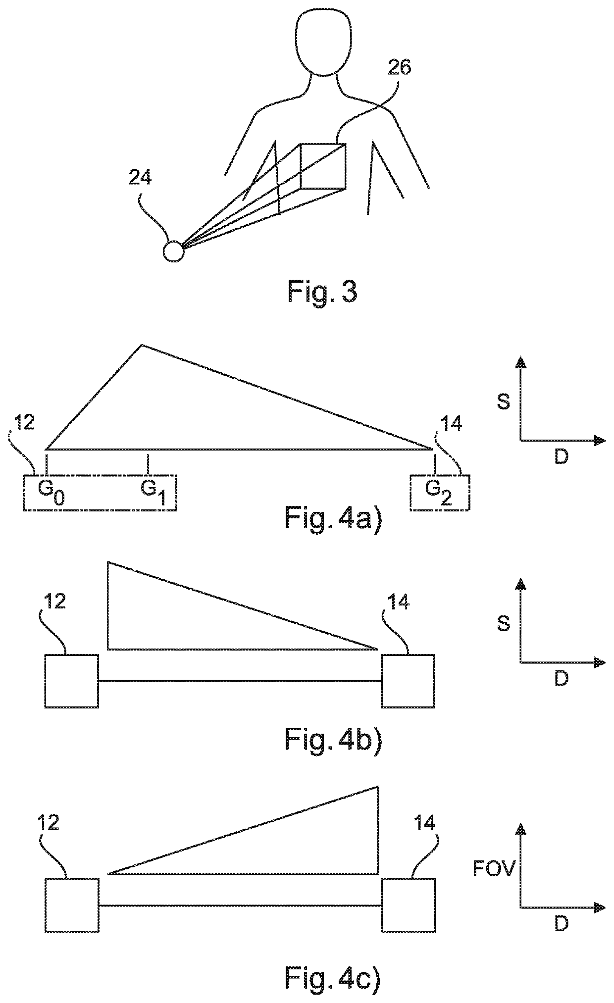
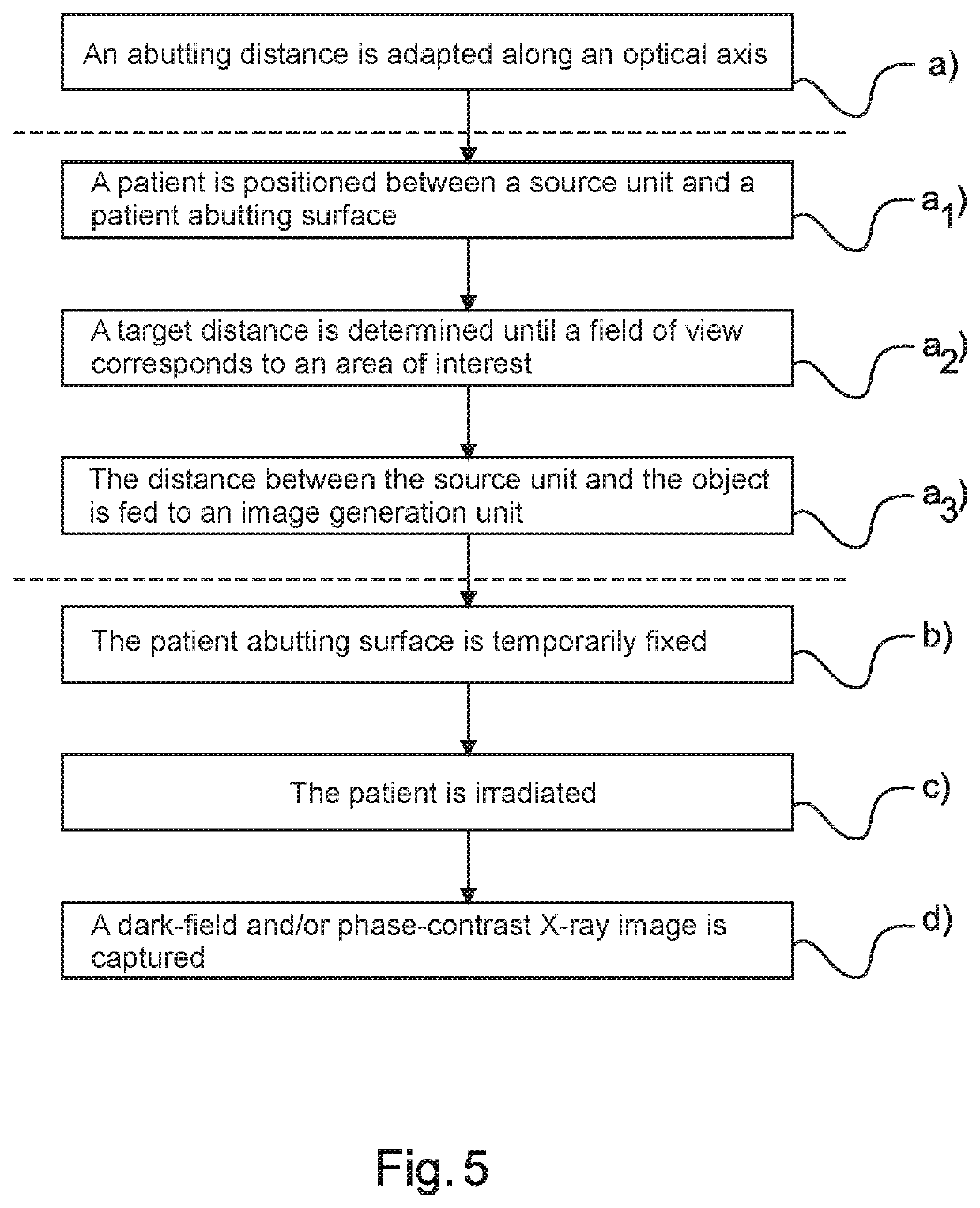
[0005]According to the present invention, a radiography system for grating based Dark-Field and / or phase-contrast X-ray imaging is provided. The radiography system for grating based Dark-Field and / or phase-contrast X-ray imaging comprises a source unit, a detection unit and a patient support with a patient abutting surface. The source unit and the detection unit are arranged along an optical axis and the patient support is arranged in between. The distance between the source unit and the patient abutting surface along the optical axis is adaptable. The abutting distance and an actual sensitivity, based on the abutting distance, are taken into account for imaging, such that a trade-off between sensitivity and field of view in a patient specific manner can be achieved.
[0006]As an effect, a high sensitivity to small angle scattering inside the object to be irradiated, i.e. the patient, is avoided, since the patient can be positioned in an adapted way. Positioning closer to the source unit of the radiation increases the magnification and thus decreases the field of view. A high sensitivity is provided as it results in a better contrast-to-noise-ratio which typically facilitates the diagnosis. Thus, the image quality is improved.
[0007]In another example, a radiography system for grating based Dark-Field and / or phase-contrast X-ray imaging is provided. The radiography system for grating based Dark-Field and / or phase-contrast X-ray imaging comprises a source unit, a detection unit and a patient support with a patient abutting surface. The source unit and the detection unit are arranged along an optical axis and the patient support is arranged in between. The distance between the source unit and the patient abutting surface along the optical axis is adaptable.

Problems solved by technology

However, it has been shown that there is a trade-off between the sensitivity of the system and the field of view that is achieved.

Method used


Image

  • Sensitivity optimized patient positioning system for dark-field x-ray imaging
  • Sensitivity optimized patient positioning system for dark-field x-ray imaging
  • Sensitivity optimized patient positioning system for dark-field x-ray imaging

Examples


Embodiment Construction

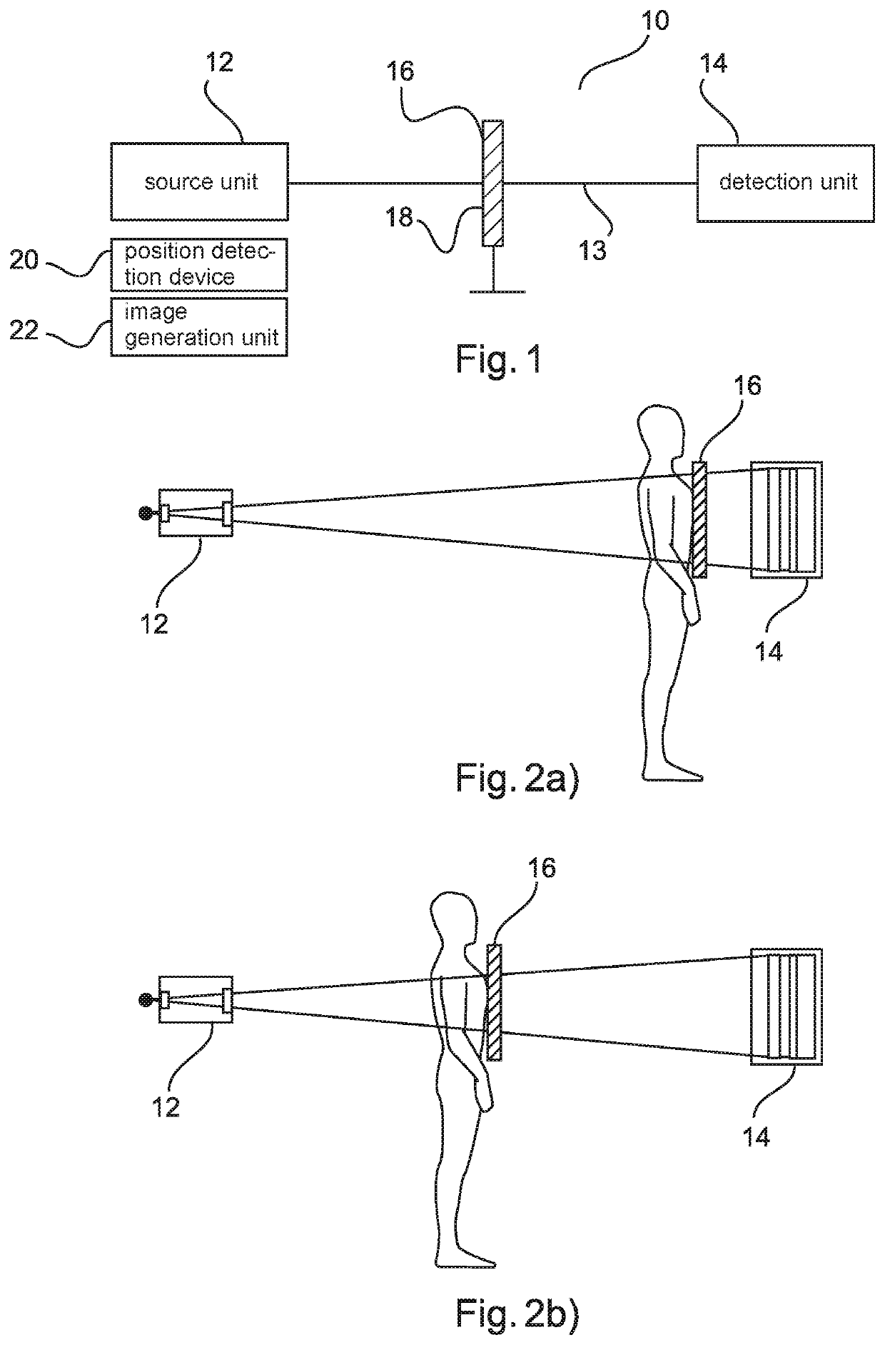
[0039]FIG. 1 shows a radiography system 10 for grating based Dark-Field and / or phase-contrast X-ray imaging. The radiography system 10 for grating based Dark-Field and / or phase-contrast X-ray imaging comprises a source unit 12, a detection unit 14 with a patient abutting surface 18. The source unit 12 and the detection unit 14 are arranged along an optical axis 13 and the patient support unit 16 with the patient abutting surface 18 is arranged in between. The patient support unit is movably arranged to be temporarily fixed in at least two different positions along the optical axis 13. The radiography system 10 for grating based Dark-Field and / or phase-contrast X-ray imaging may further comprise a position detection device 20 and an image generation unit 22. The position detection device 20 is configured to determine an actual position of the patient abutting surface 18 and to feed the actual position into the image generation unit 22, and the image generation unit 22 uses an actual ...


PUM


Abstract

The present invention relates to grating based Dark-Field and / or phase-contrast X-ray imaging. In order to improve the quality of an image, a radiography system (10) for grating based Dark-Field and / or phase-contrast X-ray imaging for imaging a patient by irradiating the patient is provided. The system comprises a source unit (12), a detection unit (14) and a patient support unit (16) with a patient abutting surface (18). The source unit (12) and the detection unit (14) are arranged along an optical axis (13) and the patient support unit (16) is arranged in between. Further, an abutting distance (dA) between the source unit (12) and the patient abutting surface (18) along the optical axis (13) is adaptable. The abutting distance (dA) and an actual sensitivity, based on the abutting distance (dA), are taken into account for imaging, such that a trade-off between sensitivity and field of view in a patient specific manner is achievable, e.g. the best trade-off.

Description

FIELD OF THE INVENTION[0001]The present invention relates to a radiography system for grating based Dark-Field and / or phase-contrast X-ray imaging and a method for capturing a Dark-Field and / or phase-contrast X-ray image.BACKGROUND OF THE INVENTION[0002]Standard X-ray imaging techniques rely on a decrease of the X-ray beam's intensity due to attenuation by an object when traversing the object to be irradiated, which can be measured with the assistance of an X-ray detector. By using interferometric methods, for instance by using a Talbot-Lau type interferometer with three gratings in the beam, two additional physical effects create contrast that can be used for imaging. Phase-contrast X-ray imaging uses information concerning changes in the phase by refraction of an X-ray beam that passes through an object in order to create image data. Dark-Field contrast X-ray imaging uses information concerning small-angle scattering. Dark-Field and / or phase-contrast X-ray imaging may take place u...

Claims


Application Information

Patent Timeline
Patent Type & Authority Patents(United States)
IPC IPC(8): A61B6/00A61B6/08
CPCA61B6/484A61B6/08A61B6/4291A61B6/589
Inventor KOEHLER, THOMASYAROSHENKO, ANDRIY
Owner KONINKLJIJKE PHILIPS NV
Learn More