Antagonistic anti-tumor necrosis factor receptor 2 antibodies
a technology of tumor necrosis factor and anti-tumor cells, applied in the field of polypeptides, can solve the problems of natural propensity in the development of this therapeutic platform, and achieve the effects of inhibiting or reducing the proliferation of cancer cells, promoting the apoptosis of cancer cells, and inhibiting or reducing the proliferation of mds
- Summary
- Abstract
- Description
- Claims
- Application Information
AI Technical Summary
Benefits of technology
Problems solved by technology
Method used
Image
Examples
example 1
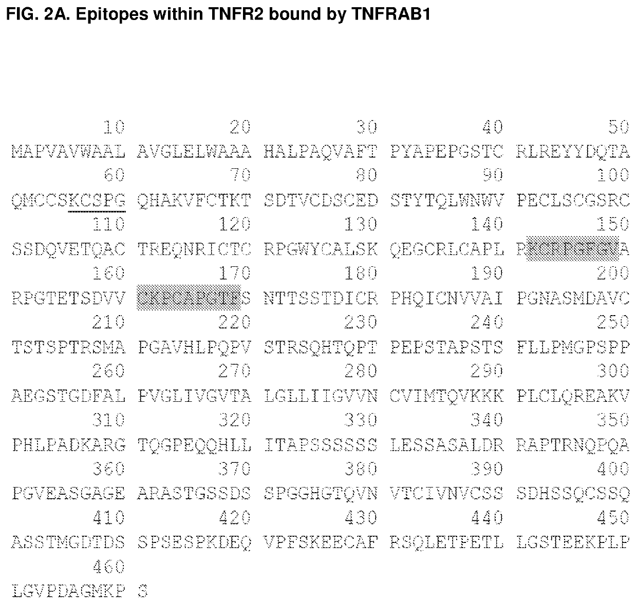
he Discrete Epitopes within TNFR2 that Interact with TNFRAB1 and TNFRAB2
[0344]Libraries of linear, cyclic, and bicyclic peptides derived from human TNFR2 were screened for distinct sequences within the protein that exhibit high affinity for TNFR2 antibody TNFRAB2. In order to screen conformational epitopes within TFNR2, peptides from distinct regions of the primary protein sequence were conjugated to one another to form chimeric peptides. These peptides contained cysteine residues at strategic positions within their primary sequences (see, e.g., FIG. 3, SEQ ID NOs: 34-117 and 134-252). This facilitated an intramolecular cross-linking strategy that was used to constrain individual peptides to a one of a wide array of three dimensional conformations. Unprotected thiols of cysteine residues were cross-linked via nucleophilic substitution reactions with divalent and trivalent electrophiles, such as 2,6-bis(bromomethyl)pyridine and 1,3,5-tris(bromomethyl)benzene, so as to form conformati...
example 2
tic TNFR2 Antibodies Inhibit T-Reg Cell Proliferation
Materials and Methods
[0352]Human T-reg Flow™ Kit (BioLegend, Cat. No. 320401)[0353]Cocktail Anti-human CD4 PE-Cy5 / CD25 PE (BioLegend, Part No. 78930)[0354]Alexa Fluor® 488 Anti-human FOXP3, Clone 259D (BioLegend, Part No. 79467)[0355]Alexa Fluor® 488 Mouse IgG1, k Isotype Ctrl (ICFC), Clone MOPC-21 (BioLegend, Part No. 79486)[0356]FOXP3 Fix / Perm Buffer (4×) (BioLegend, Cat. No. 421401)[0357]FOXP3 Perm Buffer (10×) (BioLegend, Cat. No. 421402)[0358]PE anti-human CD25, Clone: BC96 (BioLegend, Cat. No. 302606)[0359]Alexa Fluor® 488 Anti-human FOXP3, Clone 259D (BioLegend, Cat. No. 320212)[0360]PBS pH 7.4 (1×) (Gibco Cat. No. 10010-023)[0361]HBSS (1×) (Gibco Cat. No. 14175-095)[0362]FBS (heat inactivated)[0363]15 ml tubes[0364]Bench top centrifuge with swing bucket rotor for 15 ml tubes (set speed 1100 rpm or 200 g)
[0365]Antagonistic TNFR2 antibodies (TNFRAB1 and TNFRAB2) were tested for the ability to suppress the proliferation of T-...
example 3
g Antagonistic TNFR2 Antibodies by Phage Display
[0368]An exemplary method for in vitro protein evolution of antagonistic TNFR2 antibodies of the invention is phage display, a technique which is well known in the art. Phage display libraries can be created by making a designed series of mutations or variations within a coding sequence for the CDRs of an antibody or the analogous regions of an antibody-like scaffold (e.g., the BC, CD, and DE loops of 10Fn3 domains). The template antibody-encoding sequence into which these mutations are introduced may be, e.g., a naive human germline sequence as described herein. These mutations can be performed using standard mutagenesis techniques described herein or known in the art. Each mutant sequence thus encodes an antibody corresponding in overall structure to the template except having one or more amino acid variations in the sequence of the template. Retroviral and phage display vectors can be engineered using standard vector construction te...
PUM
 Abstract
Description
Claims
Application Information
 - R&D
- Intellectual Property
- Life Sciences
- Materials
- Tech Scout
- Unparalleled Data Quality
- Higher Quality Content
- 60% Fewer Hallucinations
Browse by: Latest US Patents, China's latest patents, Technical Efficacy Thesaurus, Application Domain, Technology Topic, Popular Technical Reports.
© 2025 PatSnap. All rights reserved.Legal|Privacy policy|Modern Slavery Act Transparency Statement|Sitemap|About US| Contact US: help@patsnap.com


