
Apparatus for angiographic optical coherence tomography in retina or choroid, and method for diagnosing diseases by using same

a technology of optical coherence tomography and choroid, which is applied in the field of angiographic optical coherence tomography in the retina or choroid and a method for diagnosing diseases using the same, can solve the problems of inability to accurately predict the results, the above-described conventional method is inevitably delayed in diagnosis, and the blood flow rate of the body becomes relatively insufficient, so as to achieve quick and objective measurement, improve the objectivity of microcirculatory measurement, and improve the effect of o

Active Publication Date: 2020-06-16
SEOUL NAT UNIV HOSPITAL +1
View PDF10 Cites 0 Cited by
  • Summary
  • Abstract
  • Description
  • Claims
  • Application Information

AI Technical Summary

Benefits of technology

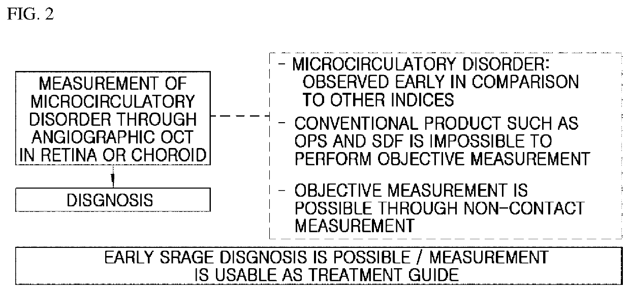
[0030]Accordingly, the angiographic optical coherence tomography apparatus of the present disclosure and the diagnostic method using the same can diagnose a shock state or a disease such as sepsis at an early stage by quickly and objectively measuring a low perfusion of tissue.
[0031]In other words, in accordance with the present disclosure, when sepsis occurs, a retina or a choroid is non-invasively and non-contactly scanned and observed using a characteristic in which a microcirculatory disorder rapidly occurs in a low perfusion of tissue, and thus sampling of the retina or the choroid is not required, so that there is an advantage in that objectivity of a microcirculatory measurement can be increased and an immediate result analysis is possible.
[0032]Further, in accordance with the present disclosure, a microcirculatory disorder can be continuously observed as well as diagnosis of disease is possible, so that there is an advantage capable of being utilized as a treatment guide through a process of observing whether a microcirculatory flow improves.

Problems solved by technology

However, the above-described conventional method is inevitably delayed in diagnosis.
When sepsis occurs, a blood flow rate of a body becomes relatively insufficient.
However, in the case of OPS and SDF imaging, a microcirculatory flow is measured at a sublingual mucosa, and there is a difference in the results depending on an observer in the microcirculatory flow measurement.
Therefore, there is a limit to objectively use OPS and SDF imaging, and thus it is necessary to develop a diagnostic technique capable of measuring a microcirculatory flow in a non-contact manner.

Method used


Image

  • Apparatus for angiographic optical coherence tomography in retina or choroid, and method for diagnosing diseases by using same
  • Apparatus for angiographic optical coherence tomography in retina or choroid, and method for diagnosing diseases by using same
  • Apparatus for angiographic optical coherence tomography in retina or choroid, and method for diagnosing diseases by using same

Examples


Embodiment Construction

[0042]Hereinafter, an apparatus for angiographic optical coherence tomography in a retina or a choroid according to the present disclosure and a disease diagnosis method using the same will be described in detail with reference to the accompanying drawings.

[0043]The present disclosure relates to an apparatus 1 for an angiographic optical coherence tomography in a retina or choroid and a disease diagnosis method using the same and relates to a diagnosis technique capable of early diagnosing a disease by quickly and objectively recognizing a low perfusion in the retina or the choroid.

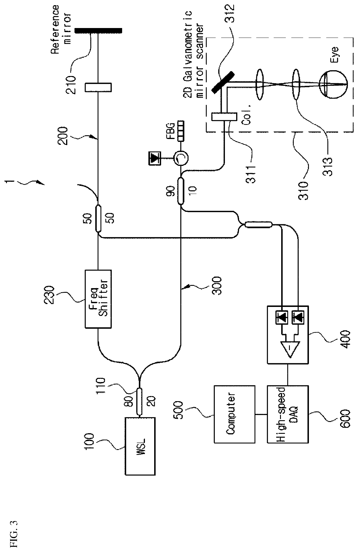
[0044]As shown in FIG. 3, the apparatus 1 for an optical coherence tomography according to the present disclosure detects an optical coherence tomography signal for a retina or a choroid and comprises a wavelength-swept laser 100, a reference arm 200, a specimen arm 300, a detector 400, and a controller 500.

[0045]In the present disclosure, the wavelength-swept laser 100 emits an optical signal to an objec...


PUM


Abstract

The present disclosure relates to an apparatus for angiographic optical coherence tomography in the retina or the choroid, and a method for diagnosing diseases by using the same and, more specifically, to: an apparatus for angiographic optical coherence tomography in the retina and the choroid, capable of diagnosing, at an early stage, shock states or diseases such as those of sepsis by quickly and objectively recognizing the low perfusion of tissue; and a diagnostic method using the same.

Description

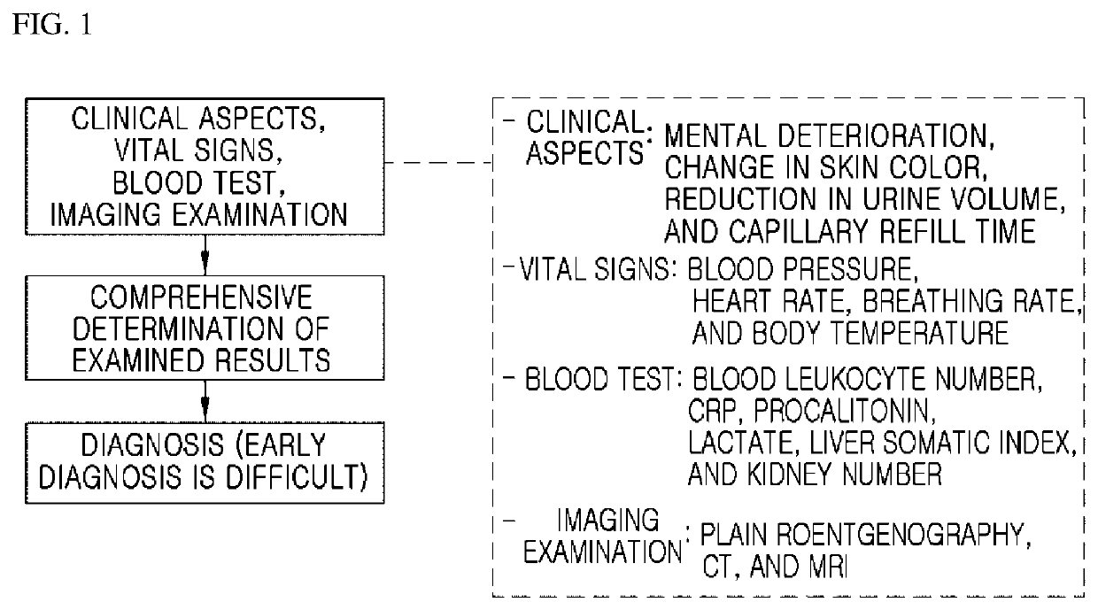
CROSS-REFERENCE TO RELATED APPLICATIONS[0001]This application is a continuation of PCT / KR2017 / 001088 filed on Feb. 1, 2017, which claims priority to Korean Patent Application No. 10-2016-0012442 filed on Feb. 1, 2016, the entire contents of which are herein incorporated by reference.FIELD OF THE INVENTION[0002]The present disclosure relates to an apparatus for an angiographic optical coherence tomography in a retina or a choroid and a method for diagnosing diseases using the same, and more particularly, to an apparatus for an angiographic optical coherence tomography in a retina or a choroid, which is capable of diagnosing a shock state or a disease such as sepsis at an early stage by quickly and objectively recognizing the low perfusion of tissue, and a diagnosis method using the same.BACKGROUND[0003]Currently, diagnosis of sepsis and shock is comprehensively determined on the basis of clinical aspects, vital signs, a blood test, imaging examination, and the like.[0004]The clinical...

Claims


Application Information

Patent Timeline
Patent Type & Authority Patents(United States)
IPC IPC(8): A61B3/14G01B9/02A61B5/00A61B3/12A61B3/10
CPCA61B5/0066A61B3/1241G01B9/02091A61B3/102A61B5/412A61B5/0035A61B3/1233G01B9/02004
Inventor OH, WANGYUHLPARK, JANG RYULKIM, KYUSEOK
Owner SEOUL NAT UNIV HOSPITAL
Learn More