
IgG subtyping assay for identifying transplantable tissue samples

a tissue sample and subtyping technology, applied in the field of organ and tissue transplantation, can solve the problems of laborious assays, insufficient indicativeness, and inability to achieve the goals of transplantation,

Active Publication Date: 2020-05-12
NJ SHARING NETWORK
View PDF3 Cites 0 Cited by
  • Summary
  • Abstract
  • Description
  • Claims
  • Application Information

AI Technical Summary

Benefits of technology

The present methods described in the patent offer the advantage of allowing multiple assessments (e.g. detection of different types of IgG) to be performed using a single sample. This avoids difficulties that can arise from reliance on multiple assays on multiple controls. Additionally, the methods use whole donor cells instead of synthetic beads to closely mimic antigen presentation by donor tissue, making the predictions more accurate. The methods were tested and shown to be highly accurate in detecting the specific type(s) of IgG that cause a positive flow cross-match, which is normally contraindicated for transplantation. The clinical implementation of this assay could increase the success rate of transplantation, especially in sensitized recipients.

Problems solved by technology

If the immune response initiated against a transplanted tissue is sufficiently severe to induce death of most or all transplanted cells or destruction of sufficient transplanted extracellular material, the transplanted tissue can fail to exhibit desirable functions or properties that were the reason for the transplant.
That is, the goals which led to the transplantation can fail to be achieved if the recipient immune response mounted against the transplanted tissue is sufficiently severe.
However, previous workers examined binding between antigen-coated beads and antibodies in recipient serum, which assays can be laborious and not necessarily indicative of interactions between recipient antibodies and donor cells.

Method used


Image

  • IgG subtyping assay for identifying transplantable tissue samples
  • IgG subtyping assay for identifying transplantable tissue samples

Examples


example 1

[0027]Isolation of PBMCs from Donor Blood

[0028]Peripheral Blood Mononuclear Cells (PBMCs, which include both lymphocytes and monocytes) were isolated from donor whole blood that was collected in acid citrate dextrose (ACD) vacutainer tubes. Donor blood was transferred to 15 milliliter conical tubes and mixed with 2 milliliters methyl cellulose (1% solution, weight / volume; Sigma-Aldrich, St. Louis, Mo., USA) by inversion. The tube was rotated in a 37 degrees Celsius incubator for 15 minutes and left standing at 37 degrees Celsius for an additional 30 minutes to separate the plasma layer containing enriched lymphocytes from the red blood cells layer. The plasma layer was carefully transferred to a fresh 15 milliliter conical tube and diluted with dulbecco's phosphate-buffered saline (DPBS, Lonza, Walkersville, Md., USA) at a ratio of 1:1 (plasma:DPBS) and mixed by inversion.

[0029]Using a Pasteur pipette, the diluted plasma was underlaid with lymphocyte separation medium (LSM) such tha...

example 2

[0034]Protocol for Identification of IgG Subtypes by Flow Cytometry

[0035]The following description is similar to the procedures described in Example 1, with some minor variations.

[0036]Isolate PBMC from whole blood or using frozen PBMC.

[0037]Wash twice with phosphate-buffered saline (PBS, pH 7.4) and perform a viability count.

[0038]Centrifuge at 1500 rpm for 5 minutes.

[0039]Discard the supernatant and resuspend the cell pellet in 0.3 ml FcR blocker reagent per 107 cells (Innovex Cat # NB309).

[0040]Incubate at room temperature (ca. 20 degrees Celsius) for 10 minutes.

[0041]Wash with excess PBS twice.

[0042]Resuspend the cells in 107 cells / ml in staining buffer (i.e., phosphate-buffered saline, pH 7.4, containing 5% v / v fetal bovine serum).

[0043]Aliquot 100 microliters per test (106 per test) of cell suspension to each 12×75 mm tube.

[0044]Add 20 microliters neat serum to each tube.

[0045]Incubate at 4 degrees Celsius for 30 minutes.

[0046]Wash with 2.5 ml staining buffer.

[0047]Discard the...

example 3

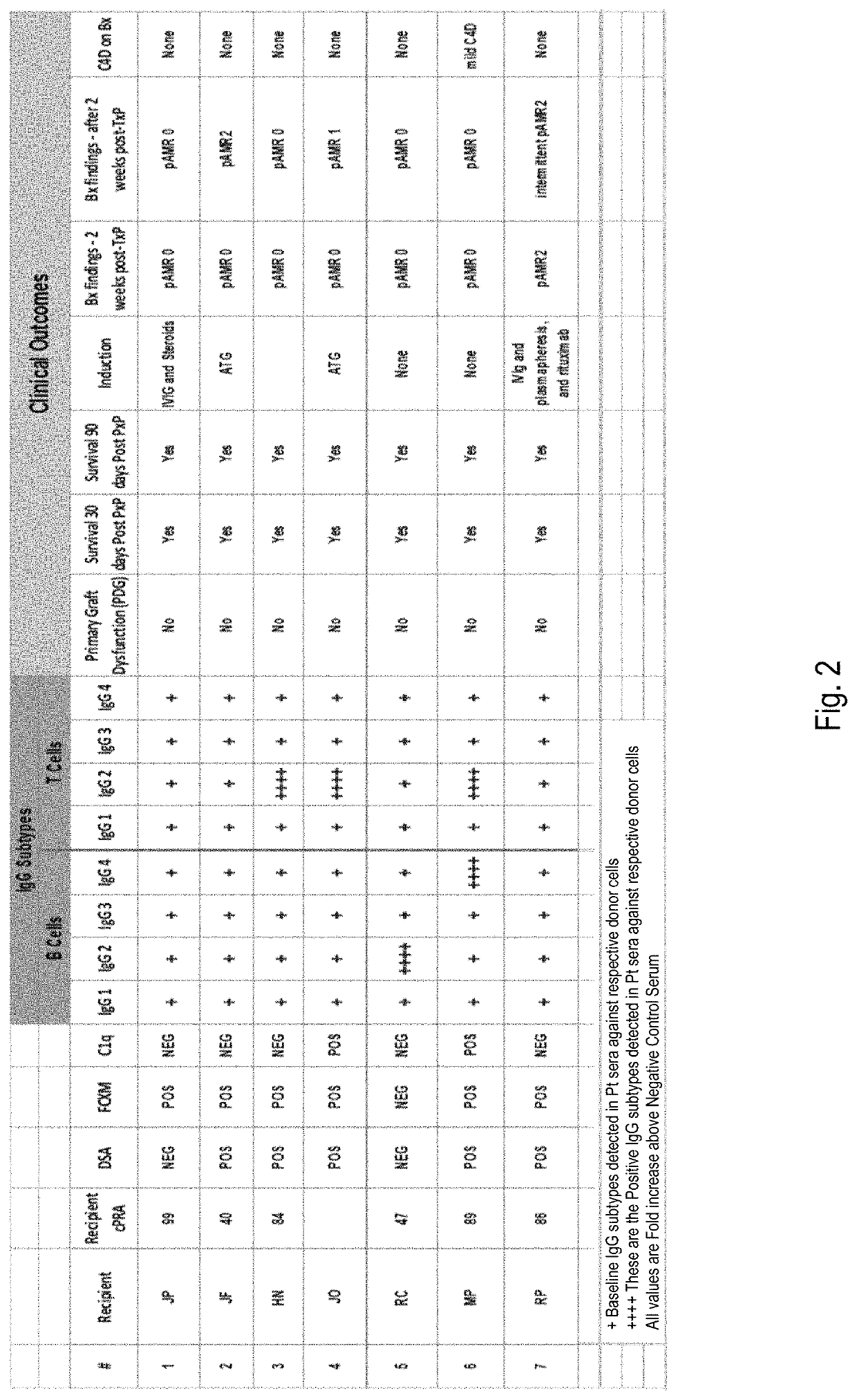
[0053]Using IgG Subtyping in Heart Transplantation Across a Positive Flow-Cytometry Cross Match (FCXM)

[0054]A positive FCXM is often a deterrent to heart transplantation due to the risk of hyperacute rejection and antibody-mediated rejection (AMR). While highly sensitive to the presence of donor-specific antibodies, FXCM does not determine whether these antibodies bind complement or not.

[0055]Case Report: 61 year old African-American male with coronary artery disease developed following myocardial infarction and coronary artery bypass graft, who developed heart failure due to ischemic cardiomyopathy with ejection fraction (EF) 15-20%. Due to progressive symptoms despite medical therapy and implantation of a cardiac resynchronization therapy device, he had a Heart Mate II left ventricular assist device (LVAD) implanted as a bridge to transplant. Post-LVAD course was complicated by recurrent gastrointestinal bleed requiring multiple transfusions as well as a driveline exit site infect...


PUM

PropertyMeasurementUnit
weight/volumeaaaaaaaaaa
volumeaaaaaaaaaa

Abstract

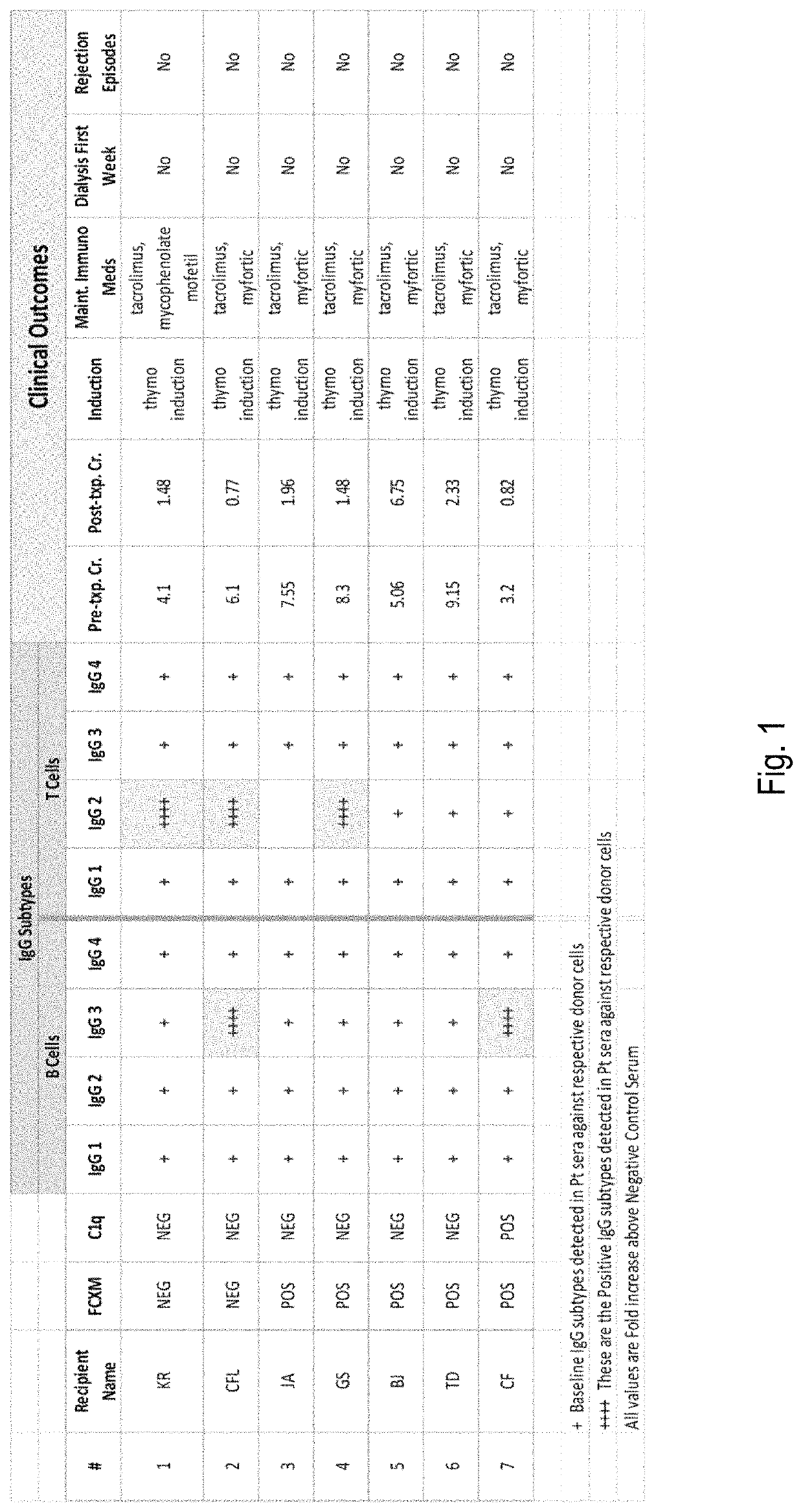
The disclosure relates to methods for assessing the suitability of a tissue obtained from a vertebrate (e.g., human) donor for grafting to a recipient, such as another vertebrate of the same species. The method involves contacting leukocytes obtained from the donor with serum from the recipient. Binding between recipient antibodies and the leukocytes is assessed, specifically focusing on the subtype(s) of IgG antibodies which bind with donor leukocytes. Detected binding between recipient antibodies of subtypes IgG1 and IgG3 indicates that the donor tissue is not suitable for grafting to the recipient. Detected binding between recipient antibodies of subtypes IgG2 and / or IgG4 indicates that the donor tissue can suitably be grafted to the recipient.

Description

CROSS-REFERENCES TO RELATED APPLICATIONS[0001]This application is entitled to priority to U.S. provisional patent application No. 62 / 262,636 filed 3 Dec. 2015.BACKGROUND OF THE DISCLOSURE[0002]The invention relates generally to the field of organ and tissue transplantation, and more specifically to procedures for matching donor tissues and recipients whose bodies will not reject the tissue if transplanted into the recipient.[0003]Transplantation of organs and tissues from the body of a donor into the body of a recipient has become a relatively common procedure. Owing to fundamental functions of the immune system of vertebrates (i.e., to alert the body to “non-self” materials and to assist in elimination of such non-self materials), tissues cannot be simply transplanted from the body of one individual of a given species into the body of any other individual of the same species without the possibility of immune complications. If the transplanted tissue is not compatible with the immun...

Claims


Application Information

Patent Timeline
Patent Type & Authority Patents(United States)
IPC IPC(8): G01N33/569G01N33/68A61K35/22A61K35/34
CPCA61K35/22G01N33/6854A61K35/34G01N33/56972G01N33/564G01N15/14G01N2015/1488
Inventor RAO, PRAKASH
Owner NJ SHARING NETWORK
Learn More